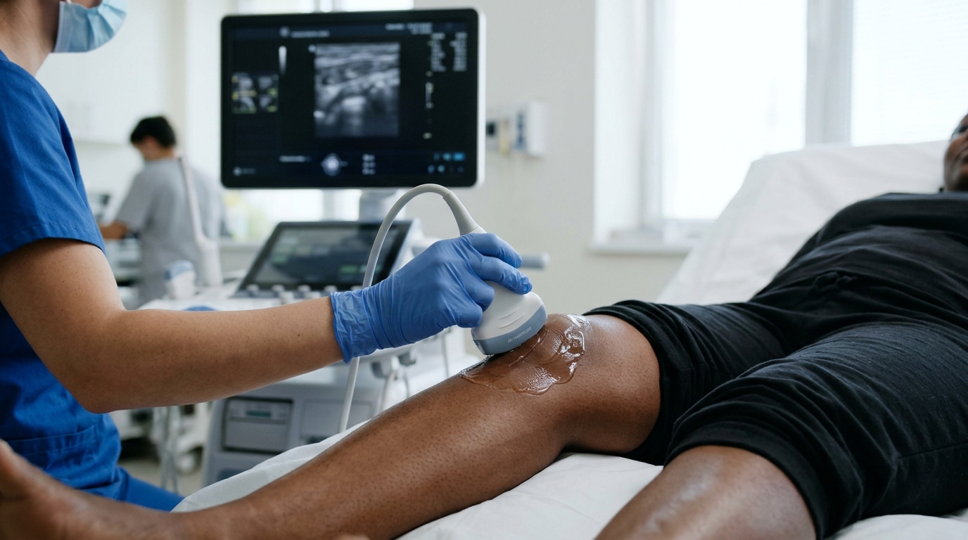
Researchers in Burkina Faso have published the first ultrasound reference values for the cartilage covering the trochlea of the knee in healthy Black African adults, based on a cross-sectional cohort of 506 participants with 1,012 knees examined at Bogodogo University Hospital. The measurements, taken with a high-frequency linear probe, showed mean cartilage thickness of 2.36 millimetres at the medial condyle, 2.38 millimetres at the lateral condyle, and 2.60 millimetres at the intercondylar zone. The work, published in BMC Rheumatology, addresses a gap the authors describe as virtually nonexistent morphometric data for sub-Saharan African populations, in contrast to the reference values available for Caucasian and Asian populations.
The participants included 408 men, or 80.63 percent of the cohort, and 98 women, or 19.37 percent, with a mean age of 24.36 years and a 95 percent confidence interval of 24.06 to 24.66 years; individual ages ranged from 19 to 45 years.
The reported mean thickness values came with confidence intervals that define the precision of the estimates: 2.36 millimetres with a 95 percent interval of 2.33 to 2.39 at the medial condyle, 2.38 millimetres with a 95 percent interval of 2.35 to 2.41 at the lateral condyle, and 2.60 millimetres with a 95 percent interval of 2.57 to 2.74 at the intercondylar zone. The intercondylar zone emerged as the thickest of the three sites on average, giving clinicians a spatial map of expected cartilage thickness across the trochlear surface rather than a single summary number.
Beyond the central values, the investigators examined which characteristics of the participants were associated with variation in cartilage thickness. In bivariate analysis, men had significantly thicker trochlear cartilage than women, with a p value of 0.001. After this multivariate adjustment, gender and body weight remained positively associated with cartilage thickness, each with a p value below 0.001, meaning that heavier participants and male participants tended to have thicker cartilage at the trochlea.
Body mass index behaved differently from body weight in the final model. While weight showed a positive association with thickness, BMI showed a negative association after adjustment. This divergence is technically meaningful because weight and BMI are related but not identical measures: BMI normalizes weight to height, so a negative BMI coefficient alongside a positive weight coefficient suggests that the two variables capture different aspects of body size in relation to cartilage thickness within this cohort. Age, which might have been expected to matter, was no longer significantly associated with thickness in the final multivariate model, a result that may reflect the narrow age range of the cohort, which spanned only 19 to 45 years with a mean in the mid-twenties.
The authors position these values as a diagnostic foundation for early detection of chondropathies in their setting, where ultrasound is the practical imaging modality for joint assessment. Reference values of this kind serve as the comparison baseline: a clinician measuring trochlear cartilage in a patient can now compare the measured thickness against values established in a population of healthy Black African adults rather than extrapolating from studies conducted on other continents. The authors also note that future research correlating these ultrasound measurements with MRI findings will help consolidate the reference values and refine the monitoring of early-stage osteoarthritis, since the present study establishes the ultrasound norms themselves rather than validating them against a second imaging modality.
The sex imbalance is pronounced, with men making up more than four-fifths of the participants, and the mean age of 24.36 years sits well below the upper limit of the 18-to-45-year recruitment range, both of which are relevant when applying the reference values to older patients or to women.
What distinguishes the Burkina Faso study is its population and its purpose: rather than testing ultrasound against another modality in diseased knees, it establishes the normal range in a population for which no such morphometric data previously existed. The authors frame the resulting dataset as an essential diagnostic foundation for their setting, and the specific numbers, 2.36 millimetres at the medial condyle, 2.38 millimetres at the lateral condyle, and 2.60 millimetres at the intercondylar zone, now stand as the reference points against which trochlear cartilage measurements in Black African adults can be interpreted.
Subject of Research: Ultrasound reference values for trochlear knee cartilage thickness in healthy Black African adults
Article Title: Ultrasound reference values for trochlear cartilage in healthy black African subjects: a study of a cohort of 506 participants
Article References: Ouédraogo, A., Nikiéma/Ouédraogo, S., Zongo, Y. E., Iname/Konate, A. S. H., Sougue, C., Coulibaly/Koudougou, J., Kambou/Tiemtoré, B. M. A., Tiendrebeogo/Zabsonré, J. W. S., & Ouédraogo, D.-D. (2026). Ultrasound reference values for trochlear cartilage in healthy black African subjects: a study of a cohort of 506 participants. BMC Rheumatology. https://doi.org/10.1186/s41927-026-00692-2
Image Credits: AI Generated
DOI: 10.1186/s41927-026-00692-2
Keywords: ultrasound, trochlear cartilage, knee, cartilage thickness, reference values, Burkina Faso, Black African adults, chondropathy, osteoarthritis, radiology, cross-sectional study, BMC Rheumatology
News Source: Ophelia Keating. (November 4, 2026). Ultrasound reference values for trochlear knee cartilage established in 506 healthy Black African adults. Scienmag.