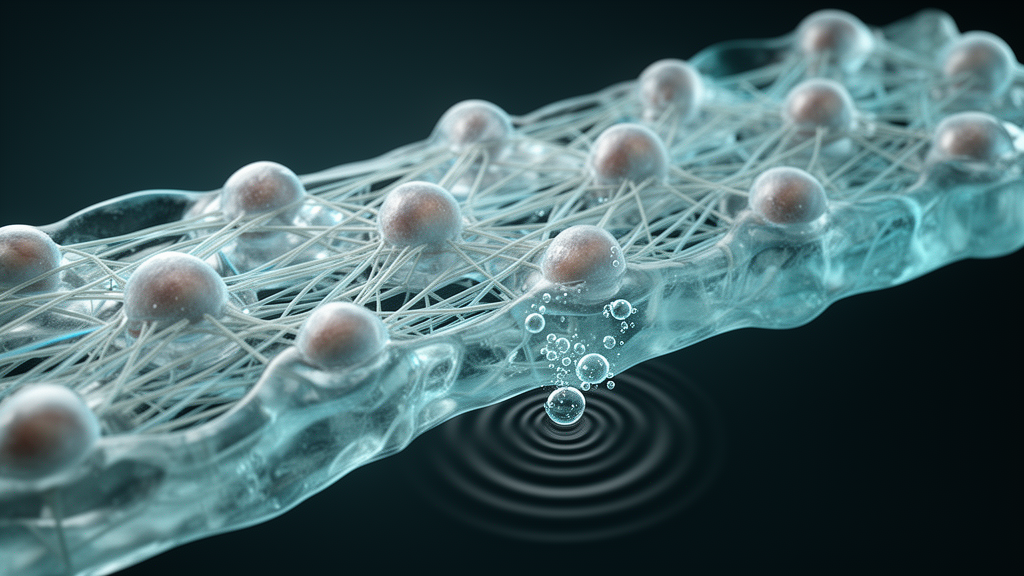
Scientists at the University of Michigan have tracked, frame by frame, what happens when tiny ultrasound-sensitive droplets vaporize into bubbles inside a tissue-like gel, revealing how far the resulting mechanical forces reach into surrounding cells. The study, published in Frontiers in Acoustics, used monodisperse perfluorohexane droplets with an average radius of about 6.35 micrometers, co-encapsulated with normal human dermal fibroblasts in fibrin hydrogels to recreate a physiologically relevant three-dimensional matrix.
The team exposed the gels to a single ultrasound burst at 2.5 megahertz, six cycles, and 5 megapascals peak negative pressure, a technique known as acoustic droplet vaporization, in which ultrasound triggers the conversion of perfluorocarbon droplets into expanding bubbles. Ultra-high-speed brightfield imaging at 10 million frames per second resolved the bubble dynamics and the displacement of cell centroids, while time-lapse confocal microscopy followed intracellular calcium signals with Fluo-4 and propidium iodide uptake for 200 seconds after the pulse.
The bubbles expanded to a maximum radius of roughly 30 micrometers, an expansion ratio of 4.7 plus or minus 1.1, and showed near-circular projections at their largest extent. Cell displacement and the inferred matrix hoop strain fell off steeply with the initial distance between a cell and the droplet. When the researchers compared their measurements with a model of incompressible spherical kinematics, evaluated per cell using each event’s maximum bubble radius, they obtained root-mean-square errors of 4.4 micrometers for displacement and 0.088 for strain. The authors note that these estimates describe matrix deformation at the cell locations rather than strain inside the cells themselves.
The spatial maps showed a clear hierarchy of effects. Hoop strains exceeded 0.1 only within about 60 micrometers of the droplet, roughly twice the maximum bubble radius, and strain rates at the bubble wall reached approximately one million per second. Propidium iodide uptake, a marker of membrane permeabilization, occurred predominantly within about 35 micrometers, close to one maximum bubble radius, where inferred hoop strains were at least 0.2.
At the boundary of the permeabilization region, cells followed two distinct trajectories: some showed a propidium iodide plateau with calcium recovery, while others displayed a continuous rise in propidium iodide together with loss of Fluo-4 signal. The researchers caution that these trajectories do not establish cell viability, and that the findings apply to the single tested exposure condition. They describe the maximum bubble radius as a useful characteristic length for the observed permeabilization region under this condition and call for further validation across other exposure parameters before cellular outcomes can be predicted more broadly. Because intracellular delivery of therapeutics is limited by the cell membrane, understanding how bubble mechanics translate into membrane permeabilization in three-dimensional matrices could inform future work on ultrasound-mediated drug delivery, though the present results remain an early mechanistic step rather than a demonstration of therapeutic efficacy.
Subject of Research: Acoustic droplet vaporization and its mechanical and cellular effects in three-dimensional fibrin hydrogels
Article Title: From bubble dynamics to cellular responses: multi-scale imaging of acoustic droplet vaporization in three-dimensional fibrin hydrogels
Article References: Chan, Z. Q., Abeid, B. A., Estrada, J. B., Fabiilli, M. L., & Aliabouzar, M. (2026). From bubble dynamics to cellular responses: multi-scale imaging of acoustic droplet vaporization in three-dimensional fibrin hydrogels. Frontiers in Acoustics, 4, Article 2003980. https://doi.org/10.3389/facou.2026.2003980
Image Credits: AI Generated
DOI: 10.3389/facou.2026.2003980
Keywords: acoustic droplet vaporization, ultrasound, perfluorohexane droplets, fibrin hydrogel, sonoporation, bubble dynamics, calcium signaling, propidium iodide, mechanobiology, fibroblasts, intracellular delivery, bubble
News Source: Denise Maddox. (October 28, 2026). Ultrasound-activated bubbles stretch cells in 3D gels, imaging study shows. Scienmag.