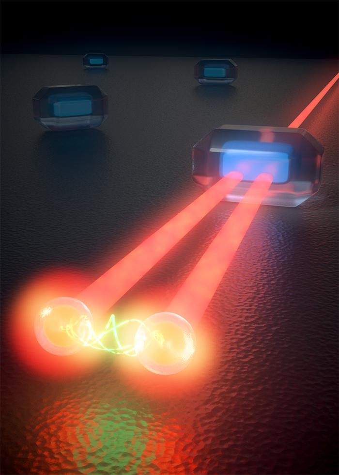
Credit: ICFO
The ability to investigate the dynamics of single particle at the nano-scale and femtosecond level remained an unfathomed dream for years. It was not until the dawn of the 21st century that nanotechnology and femtoscience gradually merged together and the first ultrafast microscopy of individual quantum dots (QDs) and molecules was accomplished. Ultrafast microscopy studies entirely rely on detecting nanoparticles or single molecules with luminescence techniques, which require efficient emitters to work. However, such techniques cause degradation to the sample, as well as, yield little information about the dynamics of the system in the excited state. Only in recent years, the efforts to find an alternative compatible technique to study fast processes in nano-objects came into the spotlight.
Now, ICFO researchers Lukasz Piatkowski, Nicolò Accanto, Gaëtan Calbris, Sotirios Christodoulou, led by ICREA Prof. at ICFO Niek F. van Hulst, in collaboration with Iwan Moreels (Ghent University, Belgium), have published a study in SCIENCE entitled “Ultrafast stimulated emission microscopy of single nanocrystals”, where they report on a technique for studying ultrafast events in individual non-fluorescent nano-objects.
In their study, they took individual QDs and rather than waiting for the QD to spontaneously emit light through photoluminescence, the team used a sophisticated combination of laser pulses to promote individual QDs into excited state and then, force them down, back to the ground state to first: image individual QDs and second: discern the evolution of the excited charges within the entire photocycle.
Dr. Lukasz Piatkowski explains why they used a laser pulse pair to effectively image the dynamics of the QDs: “It is like throwing a ball into a tree; the higher you throw it, the more excited the state. The first laser pulse of the system (photon) throws the first ball (charge in the QD) into the tree. If you are using a photoluminescence technique it is like you are standing below the tree, and you cannot see what is happening inside the treetop or crown. Thus, you will not know whether the ball starts to bounce down the branches, where, when and how is starts to fall down, if it stomps with something on its way, if it gets caught in an intermediate branch, etc. So, in order to see what is happening with the first ball, you need to find another technique that allows you to look into the treetop. The technique we used allowed us to throw a second ball into the tree top (second laser pulse interacting with the QD) to bring the first ball down. Throwing the second ball higher or lower, stronger or weaker, sooner or later after the first ball, we obtain information about the first ball and the structure of the tree (how long it took the balls to fall out, where, how, etc.) “.
In their experiment, the first laser pulse brings individual QD to the excite state. Then, every few hundreds femtosecond, they shot a second laser pulse onto the QD to bring the charges down to ground state, inducing recombination and emission of an extra photon. Hence, for every probe photon they shot into the system, they got two twin photons back. These extra photons allowed the authors not only to image the QDs but also to precisely track the evolution of the excited charges in the QD, unveiling how many charges underwent spontaneous recombination, stimulated recombination and excited state absorption.
Being able to track excited charges at the nanoscale is of fundamental importance in nanotechnology, photonics and photovoltaics. The results of the study have proven that ultrafast stimulated emission microscopy can be used to study ultrafast processes in individual chromophoric particles that are otherwise undetectable through fluorescence/photoluminescence techniques. In other words, such study has permitted imaging and studying the dynamics of nano-particles and structures without the need of external fluorescent labels.
As ICREA Prof at ICFO Niek van Hulst remarks, “Significant advances are expected in the future within the field of ultra-fast-nano-regime imaging techniques. The first detection of quantum dots using this approach has been outstanding. We now aim to extend this to molecules and biomolecular complexes, specifically photo-synthetic complexes. We are currently working on 3 and 4 pulse schemes to merge the stimulated emission and luminescence detection of single systems with 2D-spectroscopy.
###
Reference: https:/
Media Contact
Alina Hirschmann
[email protected]
0034-935-542-246


