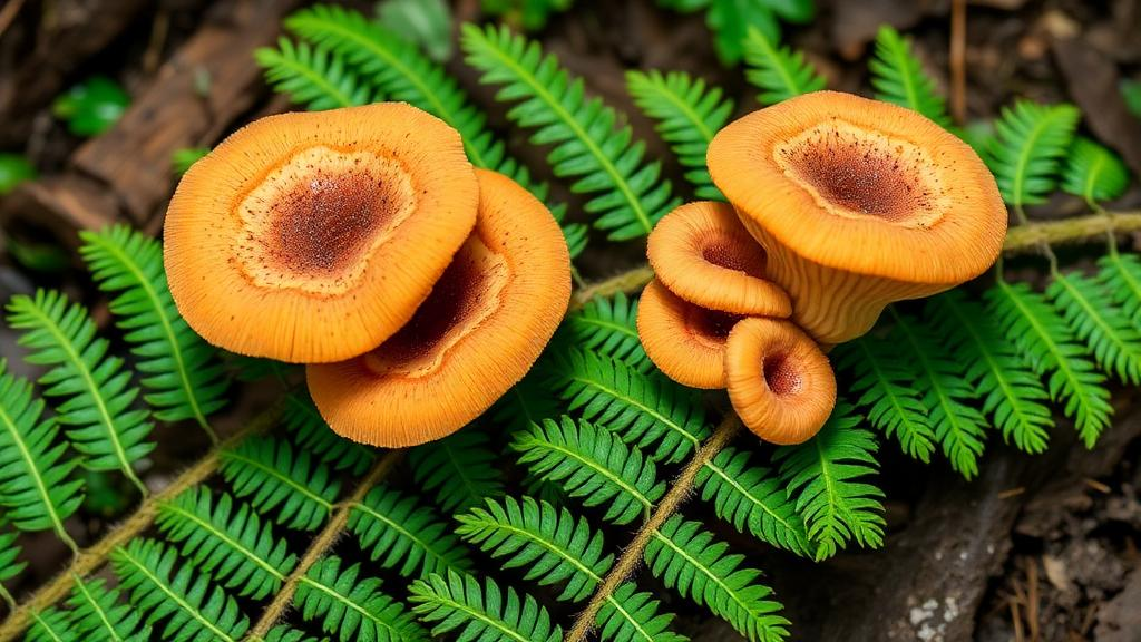
A morpho-phylogenetic survey of microfungi associated with decaying fern material in southwestern China identified two previously unknown Bionectriaceae species and documented a new host for a third fungus. The new species, Musananaesporium pronephrii and Waltergamsia yunnanensis, were recovered from dead fern stems and rachises in Guizhou and Yunnan provinces. The study also recorded Protocreopsis alba on the dead frond stalks of Angiopteris fokiensis in Guiyang. The investigators combined microscopic measurements and cultural observations with DNA sequences from four genes to classify the collections and evaluate their relationships with previously described taxa.
The three collections were obtained during a survey of terrestrial habitats, and the fungal structures developed in white, velvety colonies on their natural substrates. Under the microscope, each collection had immersed mycelium composed of branched, septate, hyaline hyphae. Their asexual forms included erect, unbranched conidiophores that tapered toward the apex and bore integrated, terminal, monophialidic conidiogenous cells. Hyaline conidia accumulated in slimy heads. Although these fungi shared a broadly similar reproductive structure, variations in conidial dimensions, form, septation and contents, together with differences in their conidiophores, provided useful features for distinguishing the taxa during the morphological examination.
For the phylogenetic work, the researchers extracted genomic DNA from fresh mycelium grown on potato dextrose agar. They amplified and sequenced the large-subunit ribosomal RNA gene region, the internal transcribed spacers, the translation elongation factor 1-alpha gene and the RNA polymerase second-largest-subunit gene. The combined matrix contained 67 taxa and 2,838 characters, including gaps, with Tilachlidium brachiatum represented by two isolates and used as the outgroup. Maximum-likelihood and Bayesian analyses produced broadly similar reconstructions. The maximum-likelihood analysis used the GTR+GAMMA model with 1,000 bootstrap repetitions, while Bayesian inference used two parallel runs of six simultaneous Markov chains for 10 million generations; the first 25% of sampled trees was excluded as burn-in.
Musananaesporium pronephrii was found on dead stems of Pronephrium penangianum, a fern in the family Thelypteridaceae, at Xianheping National Forest Park in Anlong County, Guizhou. The holotype was collected on 16 March 2022 and deposited as HKAS 129760, with the ex-type culture maintained as KUNCC 23-13971. Its specific epithet refers to the fern genus Pronephrium. The species has unbranched, septate conidiophores measuring 48–75 micrometres in length and 3–5 micrometres in width at the base. Its terminal monophialides measure 6–15 micrometres. The conidia are narrowly fusoid, straight or slightly curved, hyaline and aseptate, measuring 19–30 by 3.5–5 micrometres, with a reported mean of 24 by 4 micrometres based on 30 conidia.
The new Musananaesporium species was the second recognized member of a genus introduced in 2023 for the fungus previously called Acremonium tectonae. In the combined phylogeny, M. pronephrii and M. tectonae formed sister clades with 100% maximum-likelihood bootstrap support and a Bayesian posterior probability of 1. Morphological and genetic differences nevertheless supported recognition of the collection as a separate species. M. tectonae can have terminal and subterminal polyphialides, and its conidia may become one- to four-septate at maturity, whereas M. pronephrii has a terminal monophialide and aseptate conidia. Its conidiophores are also shorter—up to 75 micrometres compared with up to 268 micrometres—and its conidia are larger. Sequence comparisons found differences of 26 base pairs in ITS, 9 in LSU, 49 in rpb2 and 37 in tef1-alpha.
Waltergamsia yunnanensis was collected from the dead rachis of an unidentified fern in the Xishuangbanna Tropical Botanical Garden, Mengla County, Yunnan. The holotype, HKAS 129840, was gathered on 16 November 2019, and its ex-type culture was deposited as GZCC 23-0746. Its name refers to Yunnan Province. This fungus has particularly small conidiophores measuring 5–7.5 micrometres long and approximately 1 micrometre wide at the base. They are unbranched, usually carry a single basal septum and possess one integrated, terminal, monophialide. Its aseptate conidia are oblong with rounded ends or oval, measuring 1.5–2 by 0.5–1 micrometres and containing two guttules. The sexual morph was not determined.
The combined phylogenetic analysis placed W. yunnanensis within Waltergamsia and made it sister to W. alkalina, with 100% maximum-likelihood bootstrap support and a Bayesian posterior probability of 1. The comparisons also included W. maroccana. W. alkalina has conidiophores measuring 13–32.8 by 1.3–2.5 micrometres that can be unbranched or poorly branched and bear terminal and lateral phialides; its cylindrical, aseptate and eguttulate conidia measure 3–5.4 by 1.2–1.8 micrometres. W. maroccana has conidiophores measuring 10.5–21.5 by 1–2.5 micrometres, with lateral phialides sometimes arranged in two to four divergent positions at a node, and ovoid to ellipsoid conidia measuring 2.8–4 by 1.7–2.4 micrometres. The shorter conidiophores, single terminal phialide and smaller, guttulate conidia distinguished W. yunnanensis from those species.
Genetic differences also supported the separation of Waltergamsia yunnanensis. Relative to W. alkalina, it differed by 35 base pairs, including eight gaps, in a 452-base-pair ITS region; by three base pairs in an 811-base-pair tef1-alpha region without gaps; and by 10 base pairs in a 756-base-pair rpb2 region without gaps. Compared with W. maroccana, the respective differences were 62 base pairs, including 16 gaps, in a 494-base-pair ITS region; 20 base pairs in the 811-base-pair tef1-alpha region; and 72 base pairs in the 756-base-pair rpb2 region. On potato dextrose agar at room temperature, W. yunnanensis produced a colony about 30 millimetres in diameter after four weeks. The colony was superficial, dry, flat and irregularly circular, with a brown centre and white periphery.
The third collection was identified as Protocreopsis alba rather than as a new species. It grew saprobically on dead frond stalks of Angiopteris fokiensis, a member of the Marattiaceae, at the Guiyang Medicinal Botanical Garden on 13 December 2021. The specimen was deposited as HKAS 129822 and the living culture as KUNCC 23-14072. Its conidiophores reach 135 micrometres in length and are 3.5–5 micrometres wide at the base. The cylindrical conidia measure 6–10.5 by 2–3.5 micrometres, with a mean of 8 by 2.5 micrometres based on 50 conidia. They are hyaline and either aseptate or occasionally divided by one septum, with slight constriction at the septum. The researchers regarded the somewhat larger conidia and occasional septation as falling within the reported intraspecific variation of P. alba.
Molecular results confirmed the identification of the fern-associated Protocreopsis collection. In BLAST searches, its LSU and ITS sequences showed 100% similarity to P. alba, while its tef1-alpha and rpb2 sequences showed similarities of 99.89% and 99.55%, respectively. In the combined tree, isolate KUNCC 23-14072 clustered with the reference strains GMBC5357 and GMBC5358. Protocreopsis alba had previously been introduced from decaying wood in China, so its occurrence on the frond stalks of A. fokiensis constitutes a new host record. The study likewise provides the first reported fern associations for Musananaesporium and Waltergamsia.
The findings add three documented fern occurrences to a poorly sampled portion of Bionectriaceae diversity. The family comprises 50 recognized genera, and only about 20 species had previously been reported from fern substrates. Research on the taxonomy and diversity of fungi associated with ferns has remained less extensive than research on seed-plant-associated fungi, despite the ecological importance of ferns and their fungal assemblages. The three saprobic collections described here were observed on dead fern tissues, where such fungi are consistent with an association with decomposing litter. The study establishes the identities, morphological limits and phylogenetic positions of the collections, but it did not determine the sexual morphs of any of them or experimentally establish their full ecological functions. It also notes that the uncertain circumscription of Protocreopsis remains a taxonomic obstacle: the genus lacks molecular data for its type species, P. zingibericola, and a future revision may require an epitype represented by a sequenced specimen.
Subject of Research: Morphological and phylogenetic identification of Bionectriaceae fungi associated with ferns in southwestern China
Article Title: Novel Bionectriaceae ( Hypocreales ) taxa associated with ferns in southwestern China: morpho-phylogenetic description of two new species and one new host record
Article References: Zhang, J.-Y., Sun, Y.-R., Zhao, G.-L., Xie, S.-Q., Lu, Y.-Z., Xiao, Y.-P., & Liu, N.-G. (2026). Novel Bionectriaceae (Hypocreales) taxa associated with ferns in southwestern China: morpho-phylogenetic description of two new species and one new host record. MycoKeys, 136, 21-36. https://doi.org/10.3897/mycokeys.136.185538
Image Credits: AI Generated
DOI: 10.3897/mycokeys.136.185538
Keywords: Bionectriaceae, fern-associated fungi, fungal taxonomy, Musananaesporium pronephrii, Waltergamsia yunnanensis, Protocreopsis alba, Hypocreales, phylogenetics, southwestern China, fungal diversity, Novel, taxa
News Source: Roger Howard. (October 20, 2026). Two New Fungal Species Found on Ferns in Southwest China. Scienmag.