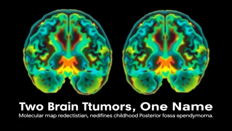
For decades, pathologists looking at ependymomas under the microscope saw one disease: a tumor of ependymal cells lining the brain’s fluid-filled ventricles, with a deceptively uniform appearance that gave little hint of how differently it could behave from one child to the next. A comprehensive new review published in the Journal of Neuro-Oncology argues that this histologic sameness has long masked a fundamental biological divide. Posterior fossa ependymomas, which account for roughly two-thirds of pediatric ependymomas and about five percent of all childhood central nervous system tumors, are in fact two molecularly distinct entities, and the distinction now shapes everything from diagnosis to surgery to radiation planning.
The review, led by George W. Koutsouras and Michael C. Dewan of Vanderbilt University Medical Center together with colleagues at the Barrow Neurological Institute, Princess Margaret Cancer Centre, and The Hospital for Sick Children, synthesizes the contemporary literature on molecular classification, imaging, and treatment outcomes for posterior fossa ependymoma. Its central message is that the World Health Organization’s shift from a purely histologic framework to molecularly integrated entities has finally caught up with the biology. Posterior fossa ependymoma group A (PFA) and group B (PFB) are not grades of the same tumor but separate diseases with different epigenetic engines, different age distributions, different imaging signatures, and sharply different prognoses.
The biological fault line runs through the epigenome. PFA tumors, which predominate in younger children with a median age of about five years, show a global loss of the repressive histone mark H3K27me3. This loss stems from inhibition of the polycomb repressive complex 2, the molecular machine that normally silences genes by trimming histone proteins, and the inhibition is commonly driven by overexpression of a protein called EZHIP, which mimics the notorious K27M oncohistone mechanism. The result is widespread derepression of developmental gene programs that fuel tumor growth. PFB tumors, by contrast, retain their H3K27me3 mark, preserve PRC2 function, and instead carry numerous arm-level chromosomal gains and losses, a pattern of somatic aneuploidy that paradoxically accompanies a more indolent clinical course. Within PFA itself, the review highlights an even more ominous stratum: tumors with chromosome 1q gain and/or 6q loss form a very high-risk group marked by increased recurrence, metastatic spread, and poor survival even after gross total resection and upfront radiotherapy.
Clinically, the two subgroups announce themselves differently. Children with posterior fossa ependymomas typically present with headache, nausea, vomiting, lethargy, and gait disturbance, the consequences of obstructive hydrocephalus as the tumor blocks cerebrospinal fluid flow from the fourth ventricle, often accompanied by truncal ataxia and sometimes lower cranial nerve deficits. Symptom onset can be insidious, and a long history of vomiting without headache may delay diagnosis. But PFA tumors tend to arise in younger children, carry a larger tumor burden, and produce more pronounced hydrocephalus, while PFB tumors appear in adolescents and adults and behave less aggressively. Even magnetic resonance imaging reflects the molecular divide: PFA tumors more often show larger volume, hydrocephalus, lateral extension through the skull base, and lower apparent diffusion coefficient values, whereas PFB tumors are typically more homogeneous, circumscribed, and cystic. The authors recommend craniospinal MRI at diagnosis, during surveillance, and at recurrence, noting that thin-cut T2-weighted spinal imaging is essential because metastatic deposits may not enhance.
When it comes to treatment, the review is unambiguous about the hierarchy of interventions. Surgery remains the single most important component of care, and the extent of resection is the key modifiable determinant of disease control. The evidence is striking. In the Children’s Oncology Group ACNS0121 study, gross total resection followed by immediate conformal radiotherapy yielded a five-year event-free survival of 68.5 percent and overall survival of 86.2 percent, compared with 37.2 percent and 70.2 percent for subtotal resection followed by adjuvant therapy. A posterior fossa-specific study by Silva da Costa and colleagues found median overall survival of 136 months after gross total resection versus 38 months after subtotal resection, with progression-free survival of 40 versus 7 months. The authors caution, however, that most surgical series are retrospective and that extent of resection partly reflects tumor anatomy, biology, and institutional experience rather than a pure treatment effect. Notably, adverse PFA biology, particularly 1q gain and 6q loss, may attenuate the survival benefit of complete resection, meaning that even a perfect operation cannot fully overcome hostile molecular terrain.
The surgical challenge itself is formidable. These tumors arise from the fourth ventricle and frequently extend through the foramina of Luschka and Magendie, wrapping around cranial nerve nuclei, the brainstem, and the vertebrobasilar circulation. In a recent molecularly defined pediatric cohort, cranial neuropathies occurred in 60 percent of patients after primary resection, though 56 percent of those resolved within a year. The review describes operative adjuncts in detail: prone positioning with individualized head fixation, since pin fixation in children under two risks calvarial injury; intraoperative neuromonitoring with spontaneous EMG recordings of cranial nerves; neuronavigation, ultrasound, and intraoperative MRI; and external ventricular drain placement, given that hydrocephalus affects up to 80 percent of children with a posterior fossa mass. When residual disease remains, early second-look surgery at an experienced center is favored, and complex ventral extension may occasionally require staged operations, always balanced against the morbidity of delaying adjuvant therapy.
Radiation therapy forms the second pillar of treatment. Postoperative focal radiotherapy is recommended for most children with localized disease after maximal safe resection, typically at a dose of 59.4 Gy, reduced to 54 Gy in selected very young patients after complete resection. Contemporary conformal techniques, including proton therapy, have progressively reduced the exposure of healthy tissue: Children’s Oncology Group protocols shrank clinical target volume margins from 10 millimeters in ACNS0121 to 5 millimeters in ACNS0831, limiting dose to normal brain and cerebellum. Proton therapy offers dosimetric advantages with disease control comparable to photon treatment, though the authors note there is no high-quality data directly comparing toxicity between modalities, and prescription doses for protons still vary from 54 to 59.4 GyE because of concerns about brainstem toxicity. Craniospinal irradiation is generally reserved for metastatic disease or recurrence, and failure after treatment remains predominantly local, underscoring that radiation efficacy depends on dose, technique, target definition, and the extent of resection achieved.
Chemotherapy, by contrast, has little to no definitive role. The randomized ACNS0831 trial found that adding four cycles of vincristine, cyclophosphamide, etoposide, and cisplatin to radiotherapy did not improve outcomes across the overall cohort, and the ACNS1021 trial showed that the targeted agent sunitinib produced no sustained objective responses. Historical baby-brain protocols that deferred radiation in children under three achieved five-year progression-free survival of at most 42 percent, while early conformal radiotherapy in comparable populations pushed seven-year progression-free survival toward 77 percent, a comparison that drove the field away from radiation-deferral strategies in children older than 12 months. One emerging exception is molecularly directed therapy: preclinical work by Griesinger and colleagues identified 1q-gain PFA as the highest-risk subgroup and demonstrated sensitivity to 5-fluorouracil and all-trans retinoic acid, a finding the review flags as a promising avenue for future investigation.
Recurrence remains the disease’s cruelest feature, occurring in up to 48 percent of cases with mortality approaching 50 percent despite multimodal therapy, and mean survival after first recurrence of roughly 31 months. More than 80 percent of recurrent infratentorial ependymomas are PFA tumors, with post-recurrence survival of 24.7 months compared with 48 months for PFB. Tumor biology evolves at relapse: 1q gain and 6q loss are enriched at recurrence, and 1q gain shortens the latency to relapse from 25.3 to 14.7 months. Recurrence can appear up to a decade after treatment, sometimes with spinal or supratentorial dissemination, which is why the authors emphasize prolonged surveillance with serial brain and spine MRI tailored to molecular risk. At relapse, management hinges on resectability: repeat surgery, even for multiply recurrent disease, is associated with improved survival, and selective re-irradiation, including craniospinal re-irradiation for posterior fossa primaries, may extend control. The review’s overall vision is a risk-adapted framework in which methylation profiling, copy number analysis, imaging biomarkers, and modern radiotherapy converge to individualize care, with treatment de-escalation for favorable PFB tumors tested only in prospective trials and molecularly directed therapies offering the clearest hope for the children whose tumors, until now, have defied every conventional weapon thrown at them.
Subject of Research: Molecular classification and management of pediatric posterior fossa ependymoma, focusing on the distinct PFA and PFB subgroups
Article Title: Posterior fossa ependymoma: a comprehensive review of molecular classification, management guidelines, and clinical outcomes (Part I of ependymomas across compartments)
Article References: Koutsouras, G. W., Rivera, F., Price, A. M., Tsang, D. S., Esbenshade, A., Ramaswamy, V., Dirks, P. B., & Dewan, M. C. (2026). Posterior fossa ependymoma: a comprehensive review of molecular classification, management guidelines, and clinical outcomes (Part I of ependymomas across compartments). Journal of Neuro-Oncology, 179(3), Article 96. https://doi.org/10.1007/s11060-026-05808-9
Image Credits: AI Generated
DOI: 10.1007/s11060-026-05808-9
Keywords: posterior fossa ependymoma, PFA, PFB, H3K27me3, EZHIP, pediatric brain tumor, gross total resection, conformal radiotherapy, proton therapy, chromosome 1q gain, 6q loss, molecular classification
Cite Scienmag News
APA MLA Chicago
Nathaniel Bowman. (September 23, 2026). Two Brain Tumors, One Name: Molecular Map Redefines Childhood Posterior Fossa Ependymoma. Scienmag. https://scienmag.com/two-brain-tumors-one-name-molecular-map-redefines-childhood-posterior-fossa-ependymoma/
Nathaniel Bowman. “Two Brain Tumors, One Name: Molecular Map Redefines Childhood Posterior Fossa Ependymoma.” Scienmag, 23 September 2026, https://scienmag.com/two-brain-tumors-one-name-molecular-map-redefines-childhood-posterior-fossa-ependymoma/. Accessed 23 September 2026.
Nathaniel Bowman. “Two Brain Tumors, One Name: Molecular Map Redefines Childhood Posterior Fossa Ependymoma.” Scienmag. September 23, 2026. https://scienmag.com/two-brain-tumors-one-name-molecular-map-redefines-childhood-posterior-fossa-ependymoma/
Copy citation Download RIS
Tags: 6q lossbrain tumor treatment planning based on molecular subtypechromosome 1q gainconformal radiotherapyEZHIPgross total resectionH3K27me3histologic versus molecular tumor diagnosisimpact of WHO tumor classification updatesimplications for surgery and radiation therapy in pediatric brain tumorsmolecular classificationmolecular classification of childhood brain tumorsmolecular mapping of ependymomaspediatric brain tumorpediatric neuro-oncology advancementspediatric posterior fossa ependymomaPFAPFA and PFB brain tumor differencesPFBposterior fossa ependymomaposterior fossa ependymoma subtypesproton therapyrole of molecular profiling in childhood CNS tumorssignificance of tumor molecular heterogeneity