The kidneys are among the most richly vascularized organs in the human body, and when clear cell renal cell carcinoma takes hold there, it exploits that blood supply with extraordinary aggression. The tumors are visibly suffused with vessels, a hallmark that has shaped decades of treatment built around blocking the growth signals that drive vessel formation. Yet patients on modern anti-angiogenic and targeted drugs often relapse, and clinicians have long lacked a convincing explanation of why a cancer so dependent on its vasculature so readily escapes therapies aimed at exactly that dependency. A new study, published in the journal Angiogenesis, offers an unusually detailed answer by looking at tumor blood vessels not as a uniform carpet of capillaries but as a heterogeneous, three-dimensional landscape containing structures that behave very differently under treatment.
The research team, led by Noémie Brassard-Jollive, Yoann Atlas, and senior authors Laurent Muller and Catherine Monnot at the Center for Interdisciplinary Research in Biology at the Collège de France, together with collaborators across Paris institutions including the Hôpital Saint-Louis pathology network, began by examining actual human ccRCC specimens at high resolution. Clear cell renal cell carcinoma is the most common form of kidney cancer and is driven overwhelmingly by loss of the von Hippel-Lindau (VHL) tumor suppressor. VHL protein normally tags hypoxia-inducible factors for destruction; without it, hypoxia signaling runs constitutively, flooding the tumor with vascular endothelial growth factor (VEGF) and other pro-angiogenic programs. The consequence is the striking hypervascularization familiar to any pathologist. But VHL loss also promotes epithelial-to-mesenchymal transition (EMT), a phenotypic switch in which epithelial tumor cells acquire invasive, migratory, fibroblast-like traits, and this connection between invasiveness and vessel architecture turned out to be central to the new findings.
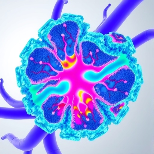
Within the patient samples, the investigators identified two vascular structures that are morphologically distinct from ordinary tumor capillaries. They named them ponds and sheets. High-resolution three-dimensional imaging of optically cleared patient-derived xenografts, essentially fragments of human tumor grown in mice and rendered transparent so that confocal microscopy could reconstruct the full vascular volume, revealed the striking geometry of these structures. Ponds appeared as large, irregular, reservoir-like formations with wide luminal cavities, vessels dilated far beyond anything resembling normal capillary caliber. Sheets, by contrast, were thin, elongated ribbons that appeared partially collapsed, as if the endothelial tubes had been flattened or incompletely organized. Neither resembles the narrow, regularly branching capillaries that pathologists count when they measure microvessel density, a metric whose prognostic value in ccRCC has proven disappointingly limited. The implication is that vessel quantity matters less than vessel architecture, and that the architecture has been largely invisible to conventional two-dimensional histology.
Capturing these structures in patients was only the first step. The team then engineered an in vitro model capable of reproducing them, because no existing laboratory system had recapitulated ccRCC vascular morphology faithfully enough to support drug testing. Their solution was a three-dimensional co-culture in which EMT-like tumor spheroids, compact spheres of tumor cells exhibiting the mesenchymal phenotype associated with VHL loss and invasion, are embedded in a dense collagen matrix alongside human endothelial cells. Collagen hydrogels of defined fibrillar density provide a physiologically relevant extracellular scaffold, and as the spheroids invade outward and the endothelial cells assemble into capillary-like networks, the two compartments interact. Crucially, the co-culture spontaneously generated structures mimicking pond architecture, whereas endothelial cells cultured alone formed conventional capillary networks. The tumor cells, in their invasive, EMT-like state, were actively instructing the formation of aberrant vascular geometry.
Time-lapse live imaging made it possible to watch this process unfold and revealed a temporal coupling that the authors describe as a link between tumor invasion and pond morphogenesis. The aberrant dilated structures did not arise independently of tumor behavior; they emerged in concert with the outward movement of invasive tumor cells, suggesting that the mechanical and biochemical traffic between invading spheroids and adjacent endothelium drives vessels into these pathological configurations. This observation ties together two features of ccRCC biology that are usually studied separately: the EMT-driven invasive program of the tumor cells and the angiogenic program of the vasculature. In this model they are two faces of a single coordinated behavior, orchestrated in part by the VHL-deficient tumor microenvironment.
The pharmacological payoff came when the team exposed the system to clinically relevant targeted therapies: temsirolimus, an mTOR inhibitor; crizotinib, a tyrosine kinase inhibitor; and sunitinib, the multi-target VEGF receptor blocker that has long anchored first-line treatment of metastatic ccRCC. In monoculture, meaning endothelial cells alone forming capillaries, and in tumor spheroid monoculture, the drugs impaired capillary morphogenesis and tumor invasion to varying degrees, consistent with known clinical activity. But when the co-culture was treated, overall drug efficacy dropped. The presence of both cell types together created a microenvironment in which the same compounds that worked well on isolated compartments became substantially less effective, an in vitro echo of the resistance that frustrates oncologists in the clinic.
The most provocative finding concerned sunitinib specifically. When the researchers analyzed the drug’s effects structure by structure within the co-culture, the pond-like formations showed markedly reduced sensitivity compared with the adjacent, more conventional capillaries. In other words, a single drug at a single concentration could silence one vascular compartment while leaving another largely intact, within the same three-dimensional culture. If ponds behave this way in patients, they would provide persistent vascular supply through the course of therapy, functioning as a structural reservoir of resistance even when standard capillaries regress. This offers a mechanistic candidate for the well-documented phenomenon of anti-angiogenic resistance in ccRCC, which has previously been attributed mostly to compensatory signaling pathways, hypoxic adaptation, and alternative pro-angiogenic factors rather than to the physical heterogeneity of the vessels themselves.
The study also carries a methodological message for the field. Two-dimensional assays, endothelial tube formation on flat Matrigel and microvessel density counts on tissue sections, dominate angiogenesis research, and the authors argue that these approaches fundamentally cannot detect structures like ponds and sheets, whose defining features are volumetric. The consensus is already shifting; recent volumetric imaging studies of ccRCC microvasculature have shown that three-dimensional vascular patterns correlate with tumor genomics and patient outcomes where density measurements fail. By combining optically cleared xenografts with a tractable in vitro co-culture that reproduces key pathological structures, the Paris team has supplied a platform on which candidate therapies can be evaluated not just for whether they reduce vessel number but for whether they eliminate the specific vascular geometries associated with invasion and drug failure.
Clinically, the identification of ponds as candidate contributors to treatment failure suggests several concrete directions. If pond formation is driven by EMT-like tumor cells, then targeting the invasive phenotype itself, or the tumor-endothelial signals that mediate pond morphogenesis, might sensitize these structures to VEGF blockade. Combination regimens pairing VEGF inhibitors with agents active against EMT-associated or mTOR-dependent pathways could be re-evaluated in the co-culture model before committing patients. And because the platform is built from human cells in a defined matrix, it could eventually support personalized testing, growing a patient’s own tumor cells in spheroid-endothelial co-culture to predict which targeted regimen will best dismantle the individual tumor’s vascular architecture.
For now, the immediate contribution is conceptual: ccRCC vasculature is not a uniform target but a heterogeneous collection of structures, some of which are intrinsically harder to drug than others, and the road to durable responses may run through the ones that pathologists’ slides have been flattening into invisibility. The study, published as volume 29, article 56 of Angiogenesis in August 2026, reorients attention from how many vessels a tumor has to what kind of vessels it has, and in doing so turns a laboratory curiosity, a dilated cavity in a collagen gel, into a plausible answer to one of kidney cancer’s most stubborn clinical questions.
Subject of Research: Aberrant three-dimensional vascular architecture (ponds and sheets) in clear cell renal cell carcinoma and its role in differential sensitivity to targeted anti-angiogenic therapies, modeled using a 3D tumor spheroid-endothelial cell co-culture system.
Subject of Research: Cancer
Article Title: 3D co-culture model of tumor spheroid and endothelial cells unveils ccRCC aberrant vasculature and distinct sensitivity to targeted treatments
Article References: Brassard-Jollive, N., Atlas, Y., Compère, C. L. M., Ardidie-Robouant, C., Mailly, P., El Bouchtaoui, M., Lelarge, V., Blot, G., Josseaume, N., De Oliveira, S., Helary, C., Leboeuf, C., Cremer, I., Sibony, M., Bousquet, G., Germain, S., Muller, L., & Monnot, C. (2026). 3D co-culture model of tumor spheroid and endothelial cells unveils ccRCC aberrant vasculature and distinct sensitivity to targeted treatments. Angiogenesis, 29(4), Article 56. https://doi.org/10.1007/s10456-026-10073-5
Image Credits: AI Generated
DOI: 10.1007/s10456-026-10073-5
Keywords: clear cell renal cell carcinoma, tumor vascularization, tumor microenvironment, 3D co-culture models, angiogenesis, anti-angiogenic therapy, endothelial cells, tumor spheroids, VHL deficiency, drug resistance, patient-derived xenografts, targeted treatment
Cite Scienmag News
APA MLA Chicago
Nathaniel Bowman. (September 8, 2026). Tumor-endothelial 3D model reveals ccRCC vessel abnormalities and drug sensitivity. Scienmag. https://scienmag.com/tumor-endothelial-3d-model-reveals-ccrcc-vessel-abnormalities-and-drug-sensitivity/
Nathaniel Bowman. “Tumor-endothelial 3D model reveals ccRCC vessel abnormalities and drug sensitivity.” Scienmag, 8 September 2026, https://scienmag.com/tumor-endothelial-3d-model-reveals-ccrcc-vessel-abnormalities-and-drug-sensitivity/. Accessed 8 September 2026.
Nathaniel Bowman. “Tumor-endothelial 3D model reveals ccRCC vessel abnormalities and drug sensitivity.” Scienmag. September 8, 2026. https://scienmag.com/tumor-endothelial-3d-model-reveals-ccrcc-vessel-abnormalities-and-drug-sensitivity/
Copy citation Download RIS
Tags: 3D tumor endothelial models3D tumor-endothelium co-culture models3D vascular modeling in ccRCCadvanced imaging of tumor blood vesselsanti-angiogenic therapy resistanceccRCC vessel abnormalitiesdrug sensitivity in renal cell carcinomahigh-resolution ccRCC tissue analysishigh-resolution ccRCC vessel imaginginterdisciplinary research in cancer biologyKidney cancer tumor-endothelial cell interactionskidney cancer vascular heterogeneitykidney tumor microenvironmentmechanisms of therapy escape in ccRCCtargeted therapy escape mechanismstumor blood vessel heterogeneitytumor microenvironment in kidney cancertumor vasculature structural abnormalitiestumor-endothelial interactionsvascular heterogeneity and treatment responsevascular structure and tumor progression