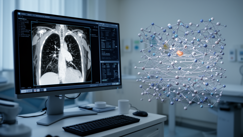
A comprehensive review of research published from 2019 through 2025 finds that transformer-based deep learning models can detect and classify lung cancer in CT and PET imaging, often outperforming conventional convolutional neural networks in internal evaluations. The findings are promising, but they do not establish a universally superior model or demonstrate clinical benefit: performance varied with the dataset, preprocessing, validation method and model design, while external testing and interpretability remained limited.
The review examined studies retrieved from IEEE Xplore, SpringerLink, ScienceDirect, PubMed, Scopus and Google Scholar. Eligible work focused on transformer methods for lung cancer detection, classification or segmentation using medical images and included peer-reviewed journal articles or conference papers with methodological developments, comparisons or experiments. Because the included studies used different populations, scanners, labels, preprocessing pipelines and validation strategies, their reported accuracy, sensitivity, specificity, area under the curve and F1 score cannot be treated as a direct league table.
Transformers adapt a language-model mechanism to images. In a Vision Transformer, or ViT, an image is divided into patches that are converted into tokens and combined with positional information. Self-attention allows each token to weigh its relationship with other tokens, enabling the model to connect a small pulmonary nodule with surrounding anatomy. A standard formulation scales the query-key product by the square root of the key dimension before applying a softmax function and weighting the values. This global interaction offers more contextual reach than the local receptive fields of CNNs, which remain computationally efficient and effective for local spatial features.
Hierarchical designs seek to retain contextual modeling with lower costs. The Swin Transformer limits attention to local windows and shifts those windows between layers so information can cross window boundaries. Reviewed studies applied Swin-based systems to nodule identification, benign-versus-malignant differentiation and tumor segmentation. Other approaches included the BiCFormer for CT classification, temporal-aware models for sequences of CT scans, 3D-NodViT for volumetric data, and hybrid CNN-transformer networks such as Swin-Tempo. The review found no architecture suited to every task: ViT was associated with strong global feature learning, Swin with more efficient hierarchical processing, and encoder-decoder systems such as UNETR and Swin-UNet with segmentation.
Reported results illustrate the potential while revealing the difficulty of comparison. One PET/CT study reported an AUC of 0.90 for malignant-lesion detection and outperformed a CNN comparator, while another reported 90% accuracy for prognosis prediction. A multimodal CT-PET system achieved 72.4% accuracy and an AUC of 0.681 for multiclass subtype classification. A Swin-plus-feature-pyramid network produced high sensitivity and specificity on LUNA16, and Swin-Tempo reported 97.84% sensitivity with 96.0% CPM. These are results from separate experiments rather than evidence of performance under the same conditions, and the review emphasizes that preprocessing, validation protocols and model choices materially affect results.
Data remain a central constraint. Public resources such as LIDC-IDRI and LUNA16 have made lung imaging research more accessible, but they differ in image quality, acquisition protocols, patient composition and annotation quality. Small nodules are especially difficult to label consistently, and benign or normal examples often outnumber malignant cases. Models trained at one institution may also encounter distribution shifts when scanners, protocols or patient populations change. The review therefore identifies standardized datasets, consistent annotation practices and multi-center collaboration as priorities for measuring whether apparent advantages survive outside the original test environment.
Computational demand is another obstacle. Global self-attention has quadratic complexity in the number of image patches, increasing memory use and training time as resolution rises, particularly for 3D CT volumes. The review discusses several ways to reduce that burden: shifted-window attention, distillation from a CNN teacher, masked-image pretraining, low-rank or sparse attention, quantization, pruning, weight sharing and low-rank adaptation of model weights. Federated learning offers a different approach, allowing institutions to train a shared model without centralizing patient data, although convergence can be slower. Lightweight variants may be better suited to shared graphics processors or edge devices, but lower resource use does not by itself establish equivalent diagnostic performance.
Interpretability and clinical workflow requirements further separate research prototypes from deployable tools. Gradient-CAM, transformer attention rollout, LIME, SHAP and saliency visualization can identify or attribute image regions involved in a prediction, but the review says relatively few studies documented these methods in detail or measured whether explanations affected trust or decisions. A model must also handle uncertainty, operate within picture archiving and communication systems, remain reliable under demographic and imaging variation, and preserve human oversight. Privacy, fairness, regulatory compliance and physician acceptance are therefore part of deployment readiness rather than separate administrative details.
The review’s conclusion is that transformer models have substantial potential for computer-aided lung cancer imaging, but current evidence supports research use more strongly than routine clinical use. A convincing next step would combine large, well-annotated multicenter datasets with transparent preprocessing, reproducible code and weights, standardized benchmarks, lightweight architectures, multimodal CT and PET information, and prospective external evaluation. Human oversight must also be tested in realistic workflows. In short, self-attention offers a powerful way to connect subtle findings with global anatomy, yet only rigorous, independent and clinically grounded validation can determine whether that technical promise translates into safer diagnosis and better care.
Subject of Research: Review of transformer-based deep learning models for lung cancer detection and classification in medical imaging.
Article Title: Transformer-based deep learning models for lung cancer detection and classification: a comprehensive review
Article References: Thakur, N., Chouksey, P., Sadotra, P., Chopra, M., Thakur, G., & Kumar, S. (2026). Transformer-based deep learning models for lung cancer detection and classification: a comprehensive review. Discover Artificial Intelligence, 6(1), Article 1090. https://doi.org/10.1007/s44163-026-02069-3
Image Credits: AI Generated
DOI: 10.1007/s44163-026-02069-3
Keywords: lung cancer, transformer models, deep learning, computer vision, CT imaging, PET imaging, medical artificial intelligence, image classification, nodule detection, clinical validation, Transformer-based, deep
News Source: Nathaniel Bowman. (October 26, 2026). Transformer Models Show Promise for Lung Cancer Imaging. Scienmag.