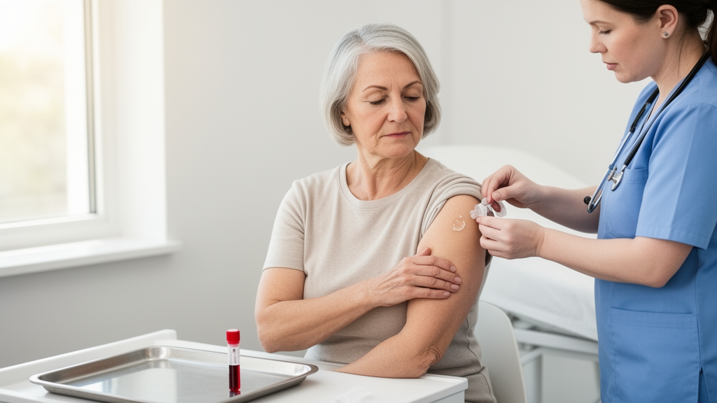
A pharmacokinetic study conducted in Japan more than two decades ago has finally been published, offering the most detailed picture yet of how a widely used transdermal estradiol gel behaves in the bodies of postmenopausal women. The open-label trial, carried out between September 1999 and January 2000 and now reported in Drugs in R&D, followed ten healthy Japanese postmenopausal women through fourteen consecutive days of once-daily application of Divigel, a 0.1% estradiol hemihydrate gel developed by Orion Pharma in Finland. The researchers measured serum estradiol and estrone, urinary excretion of three estrogens, and the pituitary hormones follicle-stimulating hormone and luteinizing hormone across three escalating doses of 0.5 mg, 1.0 mg, and 1.5 mg.
The central finding was that, with the application area standardized to approximately 400 square centimeters of thigh skin, systemic exposure to estradiol rose in proportion to the applied dose. After the final application on day 14, the time-averaged serum estradiol concentrations, calculated as the area under the concentration-time curve over 24 hours divided by the dosing interval, were 12.4 plus or minus 7.0 pg/mL at the 0.5-mg dose, 29.7 plus or minus 7.3 pg/mL at 1.0 mg, and 55.0 plus or minus 33.1 pg/mL at 1.5 mg. Steady-state estradiol concentrations were generally reached between days 3 and 5 at the two higher doses, while at the lowest dose serum levels continued to climb gradually until the last administration.
Dose proportionality was not perfectly uniform, however. At steady state, both mean maximum concentration and 24-hour exposure increased proportionally when the dose was raised from 0.5 mg to 1.0 mg, but the step from 1.0 mg to 1.5 mg produced a more than proportional increase in both parameters. The authors attribute this distortion to a single outlier subject at the 1.5-mg dose whose estradiol concentrations were markedly elevated; when median values of dose-normalized exposure were examined instead, proportionality held across all three dose levels. Individual maximum serum estradiol concentrations on day 14 ranged widely, from 13.5 to 43.3 pg/mL at 0.5 mg, 23.2 to 62.4 pg/mL at 1.0 mg, and 44.7 to 361.0 pg/mL at 1.5 mg, underscoring the considerable interindividual variability that has long been recognized with transdermal estradiol products.
The standardization of application area was not incidental. Previous work had shown that when the same estradiol dose is spread over different skin surface areas, systemic exposure and peak concentration vary inversely with the size of the area, apparently because a larger spread accelerates evaporation of the alcoholic excipient and reduces penetration. This is why approved Divigel doses are recommended to be applied over one to two palms of skin. In the present study, participants applied the gel themselves as a 20-centimeter square on either the right or left anterior thigh, on the same site as consistently as possible, a practice the authors suggest enhances saturation of estradiol in the stratum corneum and adjacent tissues.
Beyond raw estradiol levels, the study tracked the ratio of estradiol to estrone, a metabolite with substantially lower estrogenic potency that is considered a therapeutically informative index of hormone therapy. At baseline, the ratio was below 0.5, consistent with the published postmenopausal range. After application, the ratio rose clearly above 1.0 within 2 to 12 hours, peaking at 3.7 four hours after the first 1.5-mg dose and at 3.2 at the two-hour mark after the last 1.5-mg application, values approximating the premenopausal profile. It then declined back to or slightly below parity, around 1.0, within 24 hours, before the next daily application. The authors note that this roughly one-to-one ratio is typical of transdermal products and contrasts sharply with oral estradiol, where roughly 95% of the dose is metabolized in the intestine and liver to estrone and estrogen conjugates, and where reported steady-state estradiol-to-estrone ratios can fall as low as 0.17.
The urinary data added an unusual dimension, as few prior pharmacokinetic studies of transdermal estrogens have examined metabolite excretion in urine. Concentrations of estrone, estradiol, and estriol in urine increased dose dependently during the treatment period compared with levels measured 24 hours before application began. In relative terms, urinary excretion of estrone and estriol markedly exceeded that of estradiol itself at all measured time ranges and all three doses, and the estradiol-to-estrone ratio in urine was the inverse of that in serum. The cumulative urinary excretion rate, expressed as estradiol equivalent, varied linearly with the number of applications, and the estimated excretion rate at each dose was about 1%.
On the pharmacodynamic side, the gel produced the hormonal effects expected of estrogen replacement. Follicle-stimulating hormone levels were consistently lower after application at every dose compared with pre-dosing baseline values, with the effect most pronounced when measured 12 hours after the preceding application. Luteinizing hormone levels also decreased, notably at the lowest 0.5-mg dose and at the 12-hour timepoint after the higher doses, with dose-response relationships appearing steeper at that earlier measurement on day 14. Prolactin, included as a comparison and safety marker, was affected only occasionally and, if anything, decreased rather than increased relative to baseline. These reductions in gonadotropins are attributed to negative feedback by elevated serum estradiol on the hypothalamus and, for follicle-stimulating hormone, directly on the pituitary gland.
Treatment was well tolerated by all ten subjects, all of whom completed the study. No serious adverse events were observed, and no symptoms were judged definitely related to the investigational drug. The most common adverse events, judged possibly related, were leukorrhea, headache, and lower abdominal pain, with other reports including fatigue, application-site warmth, neck stiffness, and breast tenderness. One subject reported a burning sensation at the application site during the lowest-dose period, leading to an overall safety evaluation of minor safety concerns for that stage, while all other stages were judged to have no safety concerns. Skin irritation examinations found no abnormalities attributable to treatment, and there were no clinically significant changes in vital signs, laboratory parameters, or electrocardiogram recordings.
The authors place their exposure data in clinical context by reference to the widely cited effective serum estradiol range of 35 to 55 pg/mL for alleviating menopausal symptoms, particularly vasomotor symptoms. Time-averaged concentrations at the 1.0-mg and 1.5-mg doses reached well past that range, and concentrations achieved with those doses fell within the effective therapeutic range across all subjects. Notably, four of the ten women treated with 1.0 mg did not reach 35 pg/mL at any timepoint during the final 24-hour period, but all of them did so when the dose was raised to 1.5 mg, reaching that threshold at least 2 to 8 hours after the last dose. The authors also caution that serum estradiol concentration does not generally predict an individual woman’s therapeutic response or her risk of adverse outcomes, and that measurement methods themselves vary in accuracy and standardization.
Several limitations deserve emphasis. The study enrolled only ten women, all healthy Japanese postmenopausal volunteers under 65 years of age who were at least three years past menopause, without climacteric symptoms and with relatively low body mass index, a factor the authors acknowledge may have influenced exposure levels. Serum estradiol showed expected interindividual and intraindividual variability, and day 1 data at the 0.5-mg dose were evaluable in only seven subjects because three had concentrations below the assay’s quantification limit of 8 pg/mL. The trial was an open-label pharmacokinetic study commenced in 1999, before clinical trial registration became applicable in the USA and European Union for such studies, and it measured pharmacodynamic biomarkers rather than symptom outcomes. Within those bounds, the authors conclude that transdermal estradiol treatment with Divigel produced dose-proportional, clinically meaningful serum estradiol levels and estradiol-to-estrone ratios in postmenopausal women, with expected hormonal pharmacodynamic effects and good tolerability, providing systematic data intended to support individualized menopausal hormone therapy.
Subject of Research: Pharmacokinetics and pharmacodynamics of repeated transdermal estradiol gel administration in postmenopausal women
Article Title: Study on the Pharmacokinetics and Pharmacodynamics of Transdermal Estradiol Gel Administered Repeatedly in Postmenopausal Women
Article References: Sirviö, J., Ahtola-Sätilä, T., Kirjavainen, M., & Rytilä, P. (2026). Study on the Pharmacokinetics and Pharmacodynamics of Transdermal Estradiol Gel Administered Repeatedly in Postmenopausal Women. Drugs in R&D. https://doi.org/10.1007/s40268-026-00552-x
Image Credits: AI Generated
DOI: 10.1007/s40268-026-00552-x
Keywords: transdermal estradiol, Divigel, pharmacokinetics, postmenopausal women, hormone replacement therapy, estradiol-to-estrone ratio, dose proportionality, follicle-stimulating hormone, luteinizing hormone, urinary estrogen excretion, menopause, estradiol hemihydrate gel
News Source: Louis Brooks. (October 28, 2026). Transdermal Estradiol Gel Shows Dose-Proportional Blood Levels in Postmenopausal Women. Scienmag.