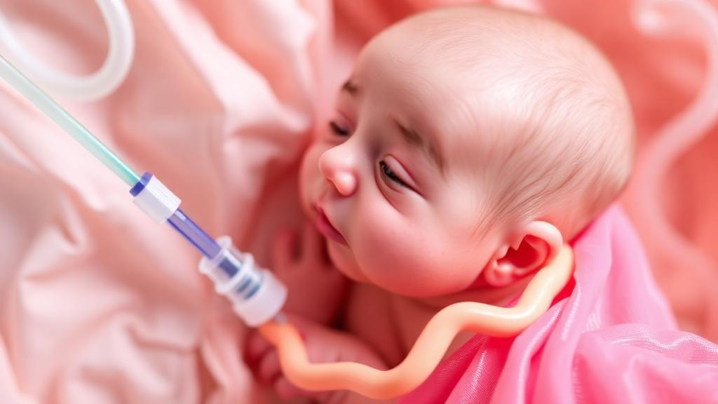
In neonatal intensive care units, the smallest patients often depend on the thinnest of medical devices. Among these are ultrathin peripherally inserted central catheters, known as PICCs, which measure just one French unit in diameter—barely a third of a millimeter. These hair-fine tubes deliver nutrition and medications directly into the central veins of preterm infants who may weigh less than a liter of milk. Yet their very thinness creates a persistent clinical problem: on a standard chest X-ray, an ultrathin catheter can be nearly invisible, leaving clinicians uncertain whether the tip has reached the safe zone of the superior vena cava or has wandered into a position that could damage tissue or trigger dangerous complications. A new retrospective observational study from Turkey now offers a practical answer, showing that injecting a small amount of iodinated contrast agent through the catheter before radiography can render the entire device clearly visible and allow precise confirmation of tip position.
The research, conducted at Adana City Training and Research Hospital and published in BMC Pediatrics, analyzed 58 preterm infants who underwent a total of 63 catheterization episodes. The cohort was extraordinarily premature, with a median gestational age of 27 weeks and a median birth weight of just 900 grams. These are babies whose skin is translucent, whose lungs are still maturing, and whose veins are fragile enough that every invasive procedure carries disproportionate risk. For this population, the authors argue, the question of catheter tip location is not a technical nicety but a matter of immediate clinical safety, because a malpositioned catheter can cause vessel perforation, cardiac arrhythmia, pleural effusion, or the inadvertent infusion of hypertonic solutions into surrounding tissue.
The technique the team evaluated is conceptually simple. Instead of relying on a plain radiograph, in which the radiopaque catheter may be indistinguishable from overlapping bones and soft tissues, clinicians instill a small bolus of iohexol—a non-ionic, low-osmolar iodinated contrast agent available at concentrations of 300 or 350 milligrams of iodine per milliliter—directly through the catheter lumen. The contrast fills the catheter and briefly opacifies its full course, so the subsequent X-ray captures not just a faint line but a sharply defined trajectory from the insertion site all the way to the tip. The authors describe a characteristic radiographic appearance they call the butterfly sign, produced when adequate opacification of the catheter creates a winged pattern at the point where the device enters the vessel. When radiographs showed superposition of structures or the absence of this butterfly sign, the team considered the image suboptimal and repeated the procedure with additional contrast.
Defining success required an anatomical standard. For catheters inserted through the upper extremities, the target tip position was set between the second and fifth thoracic vertebral levels, corresponding to the lower superior vena cava or the cavoatrial junction. For lower extremity insertions, the acceptable range was between the ninth and tenth thoracic vertebral levels, where the catheter tip should rest within the inferior vena cava. These landmarks matter because the ideal tip location balances two competing hazards: a tip that is too peripheral increases the risk of phlebitis and thrombosis, while a tip that advances too far toward or into the heart can provoke arrhythmias or cardiac perforation. In adults, tip verification is routine; in infants weighing under a kilogram, the margin of error shrinks to a few millimeters, and the bony landmarks themselves are tiny and difficult to interpret.
The results revealed how far current practice falls short of that ideal. Only 20 percent of the catheters in the study were positioned at the predefined target level on initial placement. In other words, four out of five catheters required some form of correction or repositioning before they could be considered safely located. This striking figure underscores the central argument of the paper: without a reliable visualization method, clinicians may be unknowingly operating with malpositioned devices, and the true rate of malposition in ultrathin PICCs has likely been underestimated simply because plain radiography could not detect it. Contrast-enhanced imaging, by making the catheter’s path explicit, converts a guess into a measurement.
Efficiency of the technique also emerged as a key finding. Across the 63 catheterization episodes, the team performed 71 contrast administrations in total, meaning that a single dose was sufficient in 84 percent of cases. The remaining episodes required a second administration, typically because the initial radiograph failed to demonstrate adequate opacification or because the butterfly sign was absent. For a population in which every handling event carries stress, heat loss, and infection risk, the ability to confirm tip position with one dose and one image represents a meaningful advantage over repeated plain films or blind repositioning attempts.
Safety was, understandably, the most scrutinized dimension of the study. Iodinated contrast agents are well tolerated in adults, but preterm infants have immature renal function, small circulating blood volumes, and a thyroid gland that avidly takes up iodine, raising theoretical concerns about contrast-induced nephropathy and iodine-induced thyroid dysfunction. The researchers therefore monitored serum creatinine at three time points—baseline, 48 to 72 hours after contrast administration, and again at 3 to 7 days—and evaluated kidney injury using the KDIGO classification. Among the 45 catheterization episodes that could be fully evaluated for renal outcomes, stage 1 acute kidney injury occurred in five cases, an incidence of 11.1 percent. Importantly, the authors report no allergic reactions, no catheter occlusions attributable to the contrast, and no clinically significant contrast-related thyroid dysfunction during the observation period.
It is worth interpreting the renal findings with appropriate caution, as the authors themselves do. Stage 1 acute kidney injury is common in extremely preterm infants for many reasons unrelated to contrast exposure, including hypotension, respiratory distress syndrome, patent ductus arteriosus, necrotizing enterocolitis, and exposure to nephrotoxic medications. Without a matched control group of infants who received ultrathin PICCs without contrast, the 11.1 percent figure cannot be attributed to iohexol itself. The study’s retrospective, single-center design limits causal inference, and the authors are explicit that no evident short-term safety signal emerged in this cohort rather than claiming definitive safety. Larger, ideally prospective, comparative studies would be needed to quantify any true renal or thyroid risk attributable to the procedure.
Even with those caveats, the clinical implications are substantial. Ultrathin 1 French PICCs have become increasingly popular in neonatology precisely because they suit the smallest veins of micro-preemies, allowing parenteral nutrition and long-term intravenous therapy without the complications of repeated peripheral cannulation. But their adoption has outpaced the availability of reliable verification methods, and many units still rely on plain radiographs that may simply fail to show the device. The contrast-enhanced protocol described in this study offers a low-cost, widely accessible alternative that requires no special equipment beyond the contrast agent and standard radiography already present in any neonatal intensive care unit. The butterfly sign, in particular, gives clinicians a simple visual quality check: if the sign is absent, the image is not trustworthy, and the study should be repeated rather than acted upon.
The broader lesson of the study extends beyond one catheter type or one hospital. In modern medicine, devices are often miniaturized faster than the diagnostic tools needed to monitor them, and the gap is filled by improvisation. This research demonstrates that a deliberate, protocolized approach—defining anatomical targets, standardizing contrast doses, setting explicit criteria for image adequacy, and prospectively tracking renal function—can close that gap even in the most fragile patients. For the 900-gram infant whose catheter tip sits a few millimeters from the heart, the difference between an invisible line on an X-ray and a clearly opacified trajectory may be the difference between uneventful therapy and a catastrophic complication. As neonatal intensive care continues to push the boundaries of viability at ever lower gestational ages, techniques like contrast-enhanced radiography remind us that sometimes the most impactful innovations are not new devices at all, but better ways of seeing the ones already in place.
Subject of Research: Contrast-enhanced radiography for localizing ultrathin PICC tips in preterm infants
Article Title: Contrast-enhanced radiography for localization of ultrathin (1 F) PICCs in preterm infants: a retrospective observational study
Article References: Çeki̇nmez, E. K., Gülaşı, S., Demi̇r, O., Coşkun, H., & Bulut, H. Ç. (2026). Contrast-enhanced radiography for localization of ultrathin (1 F) PICCs in preterm infants: a retrospective observational study. BMC Pediatrics. https://doi.org/10.1186/s12887-026-07573-0
Image Credits: AI Generated
DOI: 10.1186/s12887-026-07573-0
Keywords: preterm infants, PICC, contrast-enhanced radiography, iohexol, catheter malposition, neonatology, NICU, acute kidney injury, butterfly sign, radiography, very low birth weight, catheter tip localization
Ophelia Keating. (October 4, 2026). Tiny Dye Trick Reveals Hidden Catheter Errors in Preterm Babies. Scienmag.