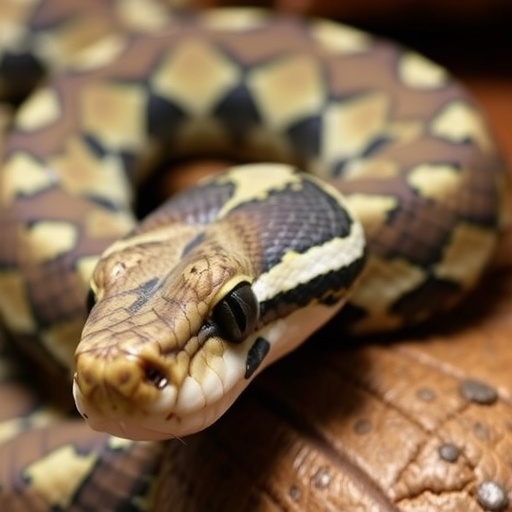
A 9-Foot Python, 754 Ticks, and Hidden Blood Parasites: Nigerian Veterinarians Rescue a Giant Snake on the Brink
When a private zoo in Abeokuta, Nigeria, called for veterinary help on 14 February 2020, the patient in question was one of the most imposing animals in their collection: an African rock python (Python sebae), roughly nine feet long and weighing 10 kilograms. But the giant constrictor was in trouble. It had refused food for an entire week, its face was visibly swollen, and its handlers reported that its skin had failed to shed properly. What veterinarians discovered during the examination—and what they painstakingly documented in a new case report published in the journal Veterinary Medicine and Science—has turned this single snake into a striking cautionary tale about parasite management in captive reptiles.
The most immediately alarming finding was on the snake’s skin. A close visual examination revealed a severe tick infestation, concentrated overwhelmingly around the head but distributed across the entire body. When the team systematically collected and preserved the ectoparasites in 70% ethanol for taxonomic identification, they counted a staggering total of 754 ticks. Of these, 382 were attached in the head region—195 nymphs, 185 adults, and just 2 larvae—while another 372, mostly adults, clung to the rest of the body under the dorsal and lateral scales. Morphological analysis under a Leica EZ4 dissecting stereomicroscope, guided by the standard taxonomic keys of Estrada-Peña and colleagues, identified every specimen as Amblyomma latum, the so-called “snake tick,” a hard tick species widespread throughout sub-Saharan Africa that frequently parasitises reptiles.
The infestation’s biology is as fascinating as it is disturbing. In A. latum, all active developmental stages may feed on a single host, and adults typically attach beneath the scales, where the head is the preferred attachment site. In captivity, the risk is compounded: confined enclosures and limited movement allow ticks to complete their entire life cycle and re-infest the same animal repeatedly. Heavy ectoparasite burdens are known to interfere with ecdysis—the delicate process of shedding—leading to dysecdysis, wounds, scarring, and deteriorating nutritional status, all of which matched the python’s clinical picture of anorexia, lethargy, and incomplete shedding.
But the ticks were only half of the story. To investigate the snake’s internal condition, the team immobilised the animal using a combination of ketamine hydrochloride at 25 mg/kg and diazepam at 0.5 mg/kg body weight, following established reptile anaesthesia protocols. They then collected blood via cardiocentesis with a sterile 23-gauge needle, preparing thin blood smears that were air-dried, fixed in absolute methanol, and stained with 10% Giemsa solution for 50 minutes. Under 1000× magnification on a Leica DM4000 B compound light microscope, the slides revealed something unexpected: dark, elongated bodies nestled inside the cytoplasm of the snake’s nucleated red blood cells.
These were intraerythrocytic gamonts—reproductive stages of haemogregarines, single-celled parasites belonging to the phylum Apicomplexa, the same broad group that includes the agents of malaria in humans. The gamonts displayed the characteristic shape, basophilic cytoplasm, and deeply stained nuclear regions consistent with the genus Haemogregarina, whose species are the most common sporozoan intracellular blood parasites of reptiles. In snakes, these parasites are typically transmitted by arthropod vectors such as leeches, mosquitoes, and ticks—or when a snake ingests an infected intermediate host such as insectivorous lizards or frogs. The authors of the report ultimately concluded that the infection represented Hepatozoon pythonis, documenting what they describe as the first recorded case of this parasite in an African rock python. Notably, A. latum itself is a known biological vector of haemoprotozoan pathogens, including haemogregarines and Hepatozoon species, raising the possibility that the tick burden and blood infection were directly linked.
Haematological analysis added further evidence of a struggle within. The packed cell volume measured 37%—well above the reference interval of 16–26% established for African rock pythons—indicating mild haemoconcentration, likely a consequence of dehydration brought on by a week of anorexia. The total white blood cell count of 27 × 10⁹ cells/L sat near the upper limit of the normal range of 7.10–29.40, hinting at subclinical infection or a stress response. Differential counts painted a more nuanced picture: heterophils at 18.10 × 10⁹ cells/L and lymphocytes at 7.83 × 10⁹ cells/L were both near their upper limits, monocytes were slightly elevated at 0.54 × 10⁹ cells/L, and eosinophils remained within normal bounds. Together, the monocytosis and borderline eosinophilia suggested a mild but genuine inflammatory response, plausibly driven by the combined assault of the haemogregarine infection and the massive tick infestation.
Treatment began immediately. The team mechanically removed the ticks, cleaned the affected areas with 10% povidone iodine, and relocated the python to a tick-free enclosure to break the re-infestation cycle. Once the anaesthesia had worn off, the snake received a single oral dose of ivermectin at 0.2 mg—a drug chosen for its documented safety in pythons and its proven efficacy against ectoparasitic infestations. The husbandry changes were just as important as the pharmacology: improved enclosure hygiene and management practices were implemented to prevent the ticks from returning.
The recovery was remarkably rapid. Within 72 hours, the python showed marked clinical improvement, and by day five it had completed a full, normal shed—clear evidence that normal ecdysis and physiological function had returned. Feeding resumed, the facial swelling resolved, and post-treatment microscopic examination of fresh blood smears confirmed the absence of Haemogregarina-like gamonts in the red blood cells, indicating that the therapeutic approach had effectively cleared the visible parasitaemia. Before-and-after images of the animal capture the transformation vividly: a swollen, tick-covered, incompletely shed snake on one side, and a sleek, healthy constrictor on the other.
The case carries lessons that extend well beyond a single snake in a Nigerian zoo. Although haemogregarine infections in reptiles are often considered apathogenic, their clinical impact can range from asymptomatic carriage to severe disease, and the true burden on host health remains poorly understood. Reptile parasites, moreover, remain understudied compared with those of mammals and birds, even though the diversity of reptilian haemoparasites likely exceeds both groups—a consequence of reptiles’ limited mobility, specialised habitats, and extraordinary evolutionary longevity. Complicating matters further, the large size and prominent nuclei of reptilian red blood cells can distort parasite morphology, making cytological identification of haemogregarine species considerably more challenging than in other vertebrates.
The authors of the report—led by researchers including Foluke Adedayo Akande and Alejandro Cabezas-Cruz—hope their findings will feed into protocols for reptile captivity and conservation programmes, and they recommend that sanitary authorities incorporate parasitic surveillance into the routine management of captive exotic reptiles. For a species that is not currently endangered but faces mounting pressure in Nigeria from habitat loss, hunting, and human-wildlife conflict, the welfare of captive populations matters as a safeguard and an educational resource. The python of Abeokuta, once crawling with more than 700 ticks and harbouring blood parasites invisible to the naked eye, recovered because of early diagnosis, targeted veterinary intervention, and a commitment to better husbandry. Its story is a vivid reminder that in the world of captive wildlife, the smallest organisms can pose the greatest threats—and that vigilance, microscopy, and rapid treatment can pull even a giant back from the brink.
Subject of Research: Severe tick infestation (Amblyomma latum) and concurrent haemogregarine (Hepatozoon pythonis) infection in a captive African rock python (Python sebae) in Nigeria
Subject of Research: Biology
Article Title: Severe Tick Infestation and Haemogregarina spp. Infection in a Captive African Rock Python (Python sebae): A Clinical and Parasitological Case from Nigeria
Article References: Akande, F. A., Diaz‑Sanchez, A. A., Egbetade, A. O., Oyenekan, I. O., Adebiyi, A. A., Oyemade, I. D., Bamgbose, T., Foucault‑Simonin, A., & Cabezas‑Cruz, A. (2026). Severe Tick Infestation and Haemogregarina spp. Infection in a Captive African Rock Python ( Python sebae ): A Clinical and Parasitological Case from Nigeria. Veterinary Medicine and Science, 12(4), Article e70571. https://doi.org/10.1002/vms3.70571
Image Credits: AI Generated
DOI: 10.1002/vms3.70571
Keywords: African rock python, Python sebae, Amblyomma latum, tick infestation, Haemogregarina, Hepatozoon pythonis, haemoparasites, captive reptiles, ivermectin, veterinary parasitology, Nigeria, reptile husbandry
Cite Scienmag News
APA
MLA
Chicago
Drew Townsend. (August 30, 2026). Ticks and Blood Parasites Strike Captive African Rock Python in Nigeria. Scienmag. https://scienmag.com/ticks-and-blood-parasites-strike-captive-african-rock-python-in-nigeria/
Drew Townsend. “Ticks and Blood Parasites Strike Captive African Rock Python in Nigeria.” Scienmag, 30 August 2026, https://scienmag.com/ticks-and-blood-parasites-strike-captive-african-rock-python-in-nigeria/. Accessed 30 August 2026.
Drew Townsend. “Ticks and Blood Parasites Strike Captive African Rock Python in Nigeria.” Scienmag. August 30, 2026. https://scienmag.com/ticks-and-blood-parasites-strike-captive-african-rock-python-in-nigeria/
Copy citation
Download RIS
Tags: African rock python parasite infestationblood parasites in reptilesblood parasites in snakescaptive reptile veterinary rescuecase report on python health crisisimpact of ectoparasites on large constrictorsimpact of parasites on snake sheddingNigeria zoo wildlife healthparasite-induced anorexia in snakesparasitic infections in captive snakesreptile health and disease managementreptile parasite identification and treatmentreptile parasite prevention in zoosreptile shedding and skin healthtick control in captive reptilestick control in zoo animalstick identification and taxonomy in reptilestick infestation in captive reptilestick infestation in captive snakesveterinary case study on Python sebaeveterinary case study on snake parasitesveterinary diagnostics for reptile infestations