For centuries, the inability of humans and other mammals to regenerate lost body parts has been viewed as an immutable biological limitation. Unlike certain amphibians such as salamanders, which can regrow entire limbs with all associated tissues intact, mammals generally heal injuries through fibrosis, a process resulting in scar tissue formation rather than true tissue regeneration. However, groundbreaking research emerging from the Texas A&M College of Veterinary Medicine and Biomedical Sciences (VMBS) challenges this long-standing paradigm. This innovative study reveals that the latent potential for regeneration may still exist in mammals, hidden within the body’s intrinsic healing mechanisms and capable of activation under specific conditions.
Aristotle pondered centuries ago why some animals could regenerate lost limbs while humans could not, a question that has captivated scientists ever since. Dr. Ken Muneoka, a distinguished professor in the Department of Veterinary Physiology & Pharmacology (VTPP) at VMBS, has dedicated his career to unraveling this mystery. His latest study, recently published in Nature Communications, showcases a novel two-step therapeutic strategy that stimulates the regeneration of bone, joint structures, and ligaments in mice digits. Although the restoration is not perfect, this method marks a vital breakthrough in approaching mammalian tissue regeneration, potentially paving the way for reduced scarring and enhanced repair following amputations.
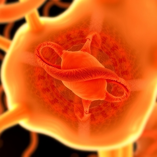
Mammalian wound healing typically induces fibrosis by activating fibroblast cells that rapidly close the injury site, forming scar tissue. This defensive response prioritizes survival but sacrifices the intricate reconstitution of complex tissues. In contrast, regenerative species like salamanders exhibit a different cellular behavior where these same fibroblast-like cells organize into a blastema. The blastema acts as a transient, specialized structure capable of orchestrating the regeneration of lost tissues. Muneoka and his team hypothesized that redirecting the behavior of fibroblasts in mammals away from scarring and towards blastema formation might unlock regenerative potential otherwise suppressed.
To test this hypothesis, the researchers designed a sequential treatment using two key growth factors: fibroblast growth factor 2 (FGF2) and bone morphogenetic protein 2 (BMP2). Critically, they timed the application of FGF2 to occur after the initial wound closure phase, allowing the body to complete its normal healing cascade before inducing a shift in fibroblast activity. The post-closure application of FGF2 stimulated the formation of a blastema-like cellular structure, an event previously thought unattainable in mammalian tissues. Several days later, BMP2 was introduced to coax these newly organized cells into differentiating and assembling into complex bone and connective tissue structures.
This two-step process embodies a sophisticated manipulation of cellular fate. The first phase steers fibroblasts away from their default scarring pathway, while the second provides instructive signals for constructing specific tissue architectures. This approach challenges the entrenched notion that regeneration requires the transplantation or supplementation of external stem cells. Instead, Muneoka stresses that the necessary progenitor cells are inherently present at the wound site; the key lies in reprogramming their behavior through targeted molecular interventions.
Dr. Larry Suva, fellow VTPP professor and co-investigator, emphasizes the paradigm shift prompted by these findings. The study reveals that cells previously assumed unprogrammable can, in fact, be redirected towards regenerative pathways. The intrinsic capacity for regeneration is not entirely lost but merely obscured by the dominant fibrotic response. Furthermore, the research uncovers evidence of positional re-specification—a phenomenon where cells can be instructed to generate structures distinct from their usual anatomical location. This discovery enriches our understanding of developmental biology and its applications in regenerative medicine.
While the regenerated digits were not precise replicas of their original anatomy, the researchers observed comprehensive restoration of critical components impaired by amputation, including bone, tendons, ligaments, and joint structures. These regenerated tissues showed organization reflecting natural biology, although some limitations in the exact form persisted. Importantly, the study highlights that mammalian regeneration involves multiple biological pathways and complex cellular coordination, refuting the oversimplified view of regeneration as a single mechanistic event.
Beyond its scientific novelty, this research holds promising translational potential. By modulating the healing process to promote regeneration rather than scarring, clinical applications could emerge to improve outcomes after injuries, surgeries, or amputations. Even partial shifts away from fibrosis could dramatically enhance tissue function, reduce chronic pain, and improve quality of life for patients. Since BMP2 is already approved by the FDA for specific medical uses and FGF2 is undergoing clinical trials, the pathway for regulatory approval of new regenerative therapies leveraging this approach may be expedited.
This study fundamentally reframes the constraints of mammalian regeneration from an absolute loss to a dormant capability waiting to be reawakened. The implications extend far beyond digit restoration, prompting investigators to explore novel questions about how regenerative processes can be harnessed and fine-tuned in humans. For Dr. Muneoka, this research lays a robust foundation upon which decades of inquiry can build new regenerative medicine paradigms, shifting from hope to tangible scientific footing.
Ultimately, this pioneering work suggests that regenerative failure in mammals is not an insurmountable barrier but a reversible state. Unlocking this latent potential could revolutionize how medicine approaches wound healing, tissue engineering, and organ restoration in the future. The research underscores the intricate interplay of cellular programming, molecular signaling, and tissue dynamics, revealing a hidden regenerative landscape within the mammalian body that science is just beginning to illuminate.
By illuminating the mechanisms that tip the balance from scar formation to tissue regrowth, the Texas A&M research team has charted a path forward for regenerative therapies poised to alter clinical practice. This landmark study invites the scientific community to reconsider the boundaries of biology and opens exciting avenues for therapeutic innovation, transforming the once-impossible dream of limb and tissue regeneration into a plausible reality.
Subject of Research: Cells
Article Title: Digit regeneration in mice is stimulated by sequential treatment with FGF2 and BMP2
News Publication Date: 17-Apr-2026
Web References:
Nature Communications Article
Texas A&M College of Veterinary Medicine and Biomedical Sciences
Image Credits: Texas A&M University
Keywords: Regeneration, Cell behavior, Fibroblast, Blastema, FGF2, BMP2, Tissue engineering, Mammalian healing, Fibrosis, Positional re-specification
Tags: amphibian limb regeneration comparisonintrinsic healing mechanisms activationjoint and ligament regeneration in miceKen Muneoka regeneration studylimb regrowth in mammalsmammalian tissue regenerationNature Communications tissue researchovercoming biological limitations in humansscar tissue versus true regenerationTexas A&M regenerative medicine researchtherapeutic strategies for bone healingveterinary regenerative science
