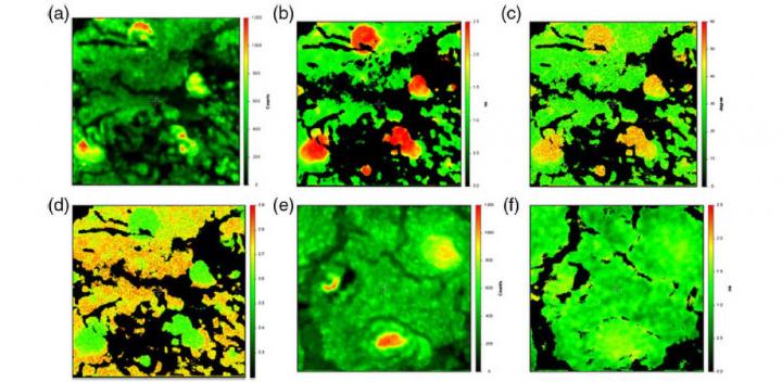
The research is published in SPIE’s Journal of Biomedical Optics

Credit: SPIE
A paper published in the Journal of Biomedical Optics (JBO), “Imaging hydroxyapatite in sub-retinal pigment epithelial deposits by fluorescence lifetime imaging microscopy with tetracycline staining,” demonstrates a potential new diagnostic option for catching a degenerative eye disease in its earliest stages.
Tiny deposits of lipids, proteins, and minerals, sometimes known as drusen, can collect under the retina. They may indicate a person’s risk of developing age-related macular degeneration (AMD), so enhanced and early detection is a current clinical need. The initial findings presented in the paper show a new way to achieve molecular contrast based upon sub-retinal mineral deposits. The approach uses a well-known property of the tetracycline-family of antibiotics – their propensity to stain teeth and bones – making visible the smallest of mineral deposits. Utilizing human cadaver retinas containing drusen, the researchers used fluorescence lifetime imaging microscopy (FLIM) to measure the light emission from tetracycline staining within those ocular mineral deposits.
According to JBO Editor-in-Chief, SPIE Fellow, and MacLean Professor of Engineering at Dartmouth Brian W. Pogue, the novel use of FLIM to harness the properties of a common antibiotic marks an exciting approach to molecular imaging in the eye, and opens up the potential for developing a diagnostic test for early transition to eye disease: “Eye diseases are typically diagnosed by shape, blood flow, or morphology changes in the retina, but this fluorescence test could be more sensitive because the signal is highly specific. It can be used to detect very small molecular mineral deposits that cannot be seen well otherwise and detect them early in the disease process. Since this is a first study, a lot more work needs to be done to make this a real human diagnostic test, but the concept that a common antibiotic could be used in this way, to reveal small mineral deposits, is a very important fundamental discovery.”
###
The article authors are Henryk Szmacinski, Department of Biochemistry and Molecular Biology, University of Maryland School of Medicine, Baltimore, Maryland; Kavita Hegde, Department of Natural Sciences, Coppin State University, Baltimore, Maryland; Hui-Hui Zeng, Department of Biochemistry and Molecular Biology, University of Maryland School of Medicine; Katayoun Eslami, Department of Biochemistry and Molecular Biology, University of Maryland School of Medicine; Adam Puche, Department of Anatomy and Neuroscience, University of Maryland School of Medicine; Imre Lengyel, Wellcome Wolfson Institute for Experimental Medicine, School of Medicine, Dentistry and Biomedical Science, Queen’s University Belfast, Belfast, UK; and Richard B. Thompson, Department of Biochemistry and Molecular Biology, University of Maryland School of Medicine.
JBO, an open-access journal, is published by SPIE in the SPIE Digital Library, which contains more than 500,000 publications from SPIE journals, proceedings, and books, with approximately 18,000 new research papers added each year.
About SPIE
SPIE is the international society for optics and photonics, an educational not-for-profit organization founded in 1955 to advance light-based science, engineering, and technology. The Society serves more than 255,000 constituents from 183 countries, offering conferences and their published proceedings, continuing education, books, journals, and the SPIE Digital Library. In 2019, SPIE provided more than $5.6 million in community support including scholarships and awards, outreach and advocacy programs, travel grants, public policy, and educational resources. http://www.
Contact:
Daneet Steffens
Public Relations Manager
[email protected]
+1 360 685 5478
@SPIEtweets
Media Contact
Daneet Steffens
[email protected]
Original Source
https:/
Related Journal Article
http://dx.


