Recent breakthroughs in terahertz (THz) wave technology are poised to revolutionize medical diagnostics by offering unprecedented insights into the microscopic architecture of biological tissues. Nestled between the infrared and microwave regions of the electromagnetic spectrum, THz waves possess unique properties that enable them to probe tissues in ways conventional imaging modalities cannot, unveiling subtle structural differences crucial for early disease detection. A new study led by Professor Hassan Arbab from Stony Brook University illuminates this potential by utilizing sophisticated mathematical models and simulations to decode how polarized THz light interacts with complex tissue environments, setting the stage for transformative advances in non-invasive medical imaging.
Traditionally, THz imaging techniques have primarily exploited contrasts based on water content differences to distinguish healthy from diseased tissues. While this approach has been somewhat effective, it falls short when confronting the intricate heterogeneity found in pathological conditions like cancer and burn injuries. The reliance on hydration levels oversimplifies tissue complexity, often masking crucial microstructural changes that could serve as reliable biomarkers of disease progression. Polarimetric measurements of THz waves—analyzing changes in wave polarization after interaction with tissue—offer a promising alternative, capable of capturing nuanced architectural features. However, the biophysical mechanisms underlying these polarization changes remained elusive until the recent computational explorations provided new clarity.
The research team harnessed Monte Carlo simulations—a statistical technique well-suited for modeling complex scattering phenomena—to explore how THz waves interact with microscopic spherical particles embedded in strongly absorbing biological media. These particles effectively represent key pathological structures found in diseased tissue, such as clusters of tumor cells or the damaged microstructures seen in burns, including the destruction of hair follicles and sweat glands. The simulations revealed that both the intensity of diffusely scattered THz light and its degree of polarization exhibit predictable variations depending on the size and concentration of these scatterers. Intriguingly, these signatures enabled the characterization of tissue polarimetric properties through a single polarization measurement, streamlining what previously demanded multiple, complex measurements.
.adsslot_wMdm0LnHsq{ width:728px !important; height:90px !important; }
@media (max-width:1199px) { .adsslot_wMdm0LnHsq{ width:468px !important; height:60px !important; } }
@media (max-width:767px) { .adsslot_wMdm0LnHsq{ width:320px !important; height:50px !important; } }
ADVERTISEMENT
Complementing their simulations, the team manufactured tissue phantoms composed of gelatin imbued with polypropylene spheres varying in size to emulate the optical properties and scattering behavior of real tissue. These experimental validations confirmed the computational predictions: larger spheres consistently yielded stronger scattered light intensity and displayed characteristic polarization dips at specific terahertz frequencies. This frequency-dependent polarimetric response sets a foundation for non-destructive, detailed tissue assessment, which could dramatically enhance diagnostic accuracy in clinical settings.
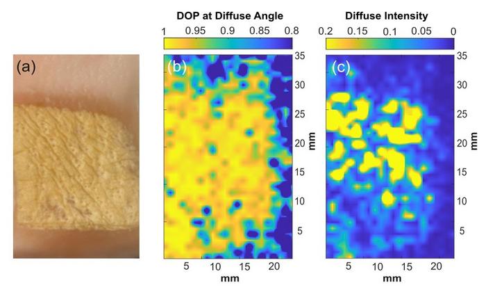
The researchers further demonstrated the clinical relevance of their approach by applying THz polarimetric imaging to porcine skin samples with induced burns, uncovering distinctive contrast between injured and healthy tissue zones. This capability suggests that THz scattering and polarimetric measurements can serve as sensitive indicators of tissue damage, holding promise for monitoring wound healing and assessing burn severity without invasive biopsies or staining—techniques currently standard in medicine but often time-consuming and resource-intensive.
Importantly, the study’s findings extend beyond burn diagnostics, offering new avenues for oncological applications. Early detection of tumor budding, where small clusters of malignant cells dissociate from the primary tumor mass, is critical for prognosis and treatment planning. Traditional detection relies on biopsy coupled with histological staining, procedures that are not only invasive but also subject to sampling errors. THz polarimetric imaging’s ability to visualize microscopic clusters through inherent tissue scattering properties presents an innovative, potentially faster diagnostic pathway, bypassing lengthy sample preparation while maintaining high sensitivity.
From a technical perspective, the study underscores the power of combining advanced computational physics with experimental optics. Monte Carlo models account for the diffuse, multiple scattering environments typical of biological tissues, a challenging scenario that hampers many conventional imaging techniques. By simulating polarized THz light’s complex interactions with tissue phantoms mimicking realistic absorption and scattering conditions, the researchers not only demystified the origins of polarimetric signals but also established quantifiable relationships between tissue microstructure and measurable optical parameters.
Looking forward, the research group plans to expand their investigations into actual cancer tissue samples, deepening the understanding of how THz polarimetric signals correlate with diverse pathological features. The development of broadband THz systems will further enable resolution of even smaller tissue structures—potentially as minute as 10 to 30 micrometers—thereby broadening the scope of detectible disease-related changes. Such advances could usher in a new paradigm of label-free, real-time tissue characterization with broad implications for early diagnosis and personalized medicine.
The implications for the medical field are profound: by offering a non-invasive, rapid, and sensitive diagnostic method, THz polarimetric imaging could reduce dependency on biopsies, lower healthcare costs, and increase patient comfort. Moreover, as THz technology matures, integration into clinical workflows might enable continuous, bedside monitoring of disease progression or therapeutic response, a feat still unachievable with many existing imaging modalities.
This study marks a significant milestone in medical optics, bridging theoretical physics, computational modeling, and experimental validation to harness the full diagnostic potential of terahertz waves. As the field moves forward, collaboration among optical physicists, engineers, and clinicians will be essential to translate these promising discoveries into effective tools for daily medical practice, potentially transforming cancer detection, burn assessment, and beyond.
In summary, the research lays out a comprehensive framework for understanding and exploiting THz Mie scattering and polarization phenomena in tissues, backed by rigorous simulation and corroborated through experimental imaging. By illuminating the subtle, yet diagnostically meaningful, variations in tissue microstructure through a novel optical window, this work sets the stage for a new generation of medical imaging technologies with remarkable sensitivity, specificity, and clinical impact.
Subject of Research: Human tissue samples
Article Title: Terahertz Mie scattering in tissue: diffuse polarimetric imaging and Monte Carlo validation in highly attenuating media models
News Publication Date: 4-Jun-2025
Web References: https://www.spiedigitallibrary.org/journals/journal-of-biomedical-optics/volume-30/issue-06/066001/Terahertz-Mie-scattering-in-tissue–diffuse-polarimetric-imaging-and/10.1117/1.JBO.30.6.066001.full
References: E. Heller et al., “Terahertz Mie scattering in tissue: diffuse polarimetric imaging and Monte Carlo validation in highly attenuating media models,” J. Biomed. Opt. 30(6), 066001 (2025). DOI: 10.1117/1.JBO.30.6.066001
Image Credits: Heller et al., doi 10.1117/1.JBO.30.6.066001
Keywords
Imaging, Oncology, Applied optics, Medical tests, Tissue damage
Tags: advanced imaging techniquesbiomarkers for disease progressionbiophysical mechanisms of polarizationburn injury detectionCancer diagnosticsmathematical models in imagingmicroscopic tissue alterationsnon-invasive medical imagingpolarized terahertz lightStony Brook University researchterahertz wave technologytissue architecture analysis


