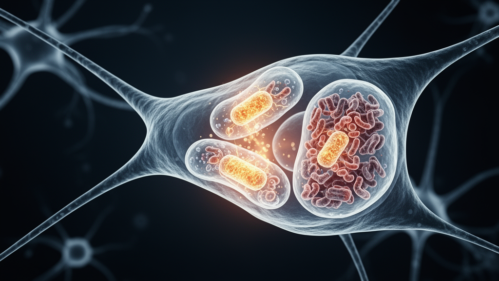
In a comprehensive scientific review published in Cell Regeneration, researchers Wenxin Li, Guo Shao, and Ruifang Qi examine how hypoxic-ischemic preconditioning orchestrates endogenous neuroprotection by modulating mitochondrial quality control and cellular bioenergetics. The authors emphasize that although the human brain accounts for only about two percent of total body weight, it consumes roughly twenty to twenty-five percent of the entire body’s oxygen supply. This exceptional metabolic demand makes neural tissue uniquely vulnerable to conditions of hypoxia and ischemia, where oxygen and nutrient deprivation rapidly impair cellular respiration. When blood supply or oxygenation drops, the brain faces immediate metabolic disruption in both neuronal and nonneuronal cells, producing neuroanatomical damage that underlies clinical conditions including acute ischemic stroke, neonatal hypoxic encephalopathy, and extreme environmental hypoxia encountered at high altitudes or in aviation. Without adequate oxygen, mitochondrial electron transport chains stall, precipitating cellular energy failure, extensive oxidative stress, neuroinflammation, and irreversible tissue damage.
Energy deficit-induced neuronal death remains an unresolved clinical challenge because effective pharmacological interventions to halt cellular necrosis remain unavailable. Although therapeutic hypothermia represents the current standard medical treatment for hypoxic-ischemic insults and reduces patient mortality to a modest extent, its therapeutic efficacy remains strictly limited. Consequently, investigators have focused on endogenous protective mechanisms triggered by hypoxic-ischemic preconditioning, an approach that applies brief, sublethal episodes of hypoxia or ischemia before a subsequent severe insult to induce tissue tolerance. While ischemic preconditioning deprives tissue of both oxygen and vital nutrients, hypoxic preconditioning relies solely on oxygen deprivation, with both paradigms mobilizing coordinated molecular cascades. Clinical investigations demonstrate that remote ischemic conditioning provides measurable benefits, such as upper-limb conditioning preventing recurrent strokes in intracranial arterial stenosis, remote conditioning reducing new ischemic events after brain tumor surgery, and similar conditioning protocols ameliorating lasting neurological deficits in ischemic moyamoya disease. These findings confirm that remote preconditioning stimulates viable systemic pathways against acute cerebral ischemia.
Mitochondria stand at the center of this endogenous defense system as the primary organelles responsible for cellular respiration and adenosine triphosphate generation. Enclosed by double membranes, mitochondria carry their own genetic material, replicate independently within host cells, and govern diverse physiological operations including calcium homeostasis, intracellular redox signaling, fatty acid oxidation, and programmed cell death. Under hypoxic and ischemic stress, mitochondrial electron transport chains and oxidative phosphorylation complexes are the first functional structures to experience disruption. This breakdown compromises cellular respiration, curtailing adenosine triphosphate synthesis and establishing a profound bioenergetic crisis. Simultaneously, the proton-motive force across the inner mitochondrial membrane declines, driving a rapid loss of mitochondrial membrane potential. In response to this electrochemical collapse, mitochondrial adenosine triphosphatase reverses its activity and rapidly hydrolyzes residual adenosine triphosphate, exacerbating cellular energy depletion and setting off irreversible structural degradation, membrane permeability changes, and ultimate necrotic cell death. Protecting these mitochondrial processes is therefore imperative to prevent extensive neurological impairment.
The review demonstrates that hypoxic-ischemic preconditioning preserves cellular energy pools by sustaining adenosine triphosphate synthesis and attenuating energy demand. Experimental investigations indicate that hypoxic preconditioning increases adenosine triphosphate concentrations in neonatal rat hippocampal tissue damaged by propofol neurotoxicity and improves hypoxia tolerance in cultured HT22 hippocampal cells. Remote ischemic preconditioning has likewise been shown to ameliorate high-altitude hypoxia-induced brain edema in animal models by rescuing the mitochondrial membrane potential and promoting tricarboxylic acid cycle glucose metabolism to augment adenosine triphosphate output. On an enzymatic level, metabolic stress alters the intracellular AMP-to-ATP ratio, triggering the energy sensor AMP-activated protein kinase. Activated AMP-activated protein kinase promotes catabolic pathways to produce adenosine triphosphate while downregulating energy-consuming anabolic processes. In ischemic brain injury models, intermittent hypoxia activates AMP-activated protein kinase, peroxisome proliferator-activated receptor gamma coactivator-1alpha, and sirtuin-3 signaling cascades, repairing mitochondrial ultrastructure and restoring cellular bioenergetic stability. Furthermore, sirtuin enzymes like sirtuin-1 activate sirtuin-3 to mitigate cerebral ischemia-reperfusion damage and reduce neuroinflammation.
A pivotal component of preconditioning-mediated neuroprotection involves regulating reactive oxygen species and maintaining physiological redox balance within neural tissue. Under normal conditions, the mitochondrial electron transport chain generates basal levels of superoxide anions that rapidly convert into hydrogen peroxide, functioning as essential secondary messengers in cellular signaling—a state known as oxidative eustress. However, severe ischemia and reperfusion overload the respiratory chain, producing massive surges of mitochondrial reactive oxygen species that cause oxidative distress. These uncontained reactive species oxidize cellular lipids, structural proteins, and nucleic acids, damaging mitochondrial integrity and triggering apoptosis. Hypoxic-ischemic preconditioning mitigates this destructive cascade by boosting endogenous antioxidant enzymes like superoxide dismutase, suppressing excessive radical production, and alleviating reperfusion injury. Furthermore, preconditioning upregulates hypoxia-inducible factor-1alpha, which elevates the expression and activity of glucose transporters GLUT1 and GLUT3, thereby stabilizing intracellular glucose availability and fueling neuronal metabolism under stress. This dual action protects astrocytes and neurons alike from lethal oxidative damage during reperfusion.
Disruption of mitochondrial function during hypoxia-ischemia also provokes pathological calcium overload, another primary driver of intrinsic neuronal death that preconditioning effectively counters. Neurons require rigorous calcium regulation to propagate action potentials, regulate excitability, and direct gene transcription, relying on mitochondria as central buffers of intracellular calcium homeostasis. When hypoxia impairs bioenergetic systems, cytosolic free calcium levels surge, overwhelming mitochondrial calcium uniporters and driving excessive calcium uptake into the mitochondrial matrix. This intra-organelle calcium overload triggers the opening of the mitochondrial permeability transition pore, depolarizing the inner membrane and causing osmotic swelling and caspase cofactor release. The review highlights that hypoxic-ischemic preconditioning prevents this collapse by modulating calcium transport proteins, specifically by upregulating sodium-calcium exchangers NCX1 and NCX3 in brain structures such as the dentate gyrus. Preserving ionic equilibrium thereby prevents cytochrome c release into the cytoplasm.
The preservation of mitochondrial membrane integrity directly attenuates the mitochondria-mediated intrinsic apoptotic pathway, which plays an essential role in post-ischemic brain damage. Intrinsic apoptosis is governed by the B-cell lymphoma 2 family of proteins, where the ratio between pro-survival proteins like Bcl-2 and pro-apoptotic executioners like Bax determines cellular fate. Under hypoxic-ischemic insults, mitochondrial swelling, loss of membrane potential, and oxidative stress induce apoptosome assembly and caspase activation. Research shows that ischemic preconditioning reduces the Bax-to-Bcl-2 ratio and suppresses caspase-3 activation, significantly limiting cerebral infarction volume and hippocampal tissue loss. Preconditioning also engages broad anti-apoptotic signaling cascades, including erythropoietin-mediated activation of phosphatidylinositol 3-kinase and extracellular signal-regulated kinase pathways, Notch1 and nuclear factor-kappa B signaling, and cyclic adenosine monophosphate-mediated phosphorylation of CREB. Together, these coordinated signaling networks suppress pro-apoptotic gene transcription, promote microglial polarization, and shield neuronal populations from delayed apoptotic destruction. Additionally, remote preconditioning stimulates mitochondria-derived vesicles to alleviate reperfusion injury.
When structural mitochondrial damage occurs despite early antioxidant defenses, cells rely on selective macroautophagy, termed mitophagy, to sequester and degrade dysfunctional organelles before they trigger apoptosis. Mitophagy operates primarily through two distinct molecular routes: the ubiquitin-mediated pathway orchestrated by PTEN-induced kinase 1 and Parkin, and receptor-mediated pathways utilizing proteins including BNIP3, NIX, and FUNDC1. The review outlines evidence showing that preconditioning enhances mitophagy to eliminate permeabilized mitochondria and protect neural cells from ischemia-reperfusion injury. In adult rat models of transient global cerebral ischemia, hypoxic preconditioning stimulates the PTEN-induced kinase 1 and Parkin ubiquitination cascade, accelerating damaged organelle clearance. Similarly, hypoxically preconditioned mesenchymal stem cell-derived extracellular vesicles activate p62-dependent mitophagy pathways, boosting adenosine triphosphate production and repairing retinal progenitor cells. This timely removal of fragmented mitochondria prevents cellular energy failure and downstream death.
Mitochondrial quality control also depends on the dynamic equilibrium between mitochondrial biogenesis, fusion, and fission, which continuously remodel the cellular mitochondrial architecture. Because eukaryotic cells cannot produce mitochondria de novo, preexisting organelles undergo balanced fusion—governed by mitofusins 1 and 2 and optic atrophy 1—to exchange contents and maintain metabolic capacity, while dynamin-related protein-1 and mitochondrial fission protein 1 mediate fission to segregate damaged segments. Hypoxic-ischemic stress disrupts this balance by promoting excessive dynamin-related protein-1 translocation, causing pathological mitochondrial fragmentation and neurovascular breakdown. Preconditioning counteracts excessive fission by suppressing dynamin-related protein-1 activation and promoting mitofusin-dependent fusion. Concurrently, preconditioning stimulates mitochondrial biogenesis through AMP-activated protein kinase and nuclear respiratory factors, driving organelle replication. In experimental models, ischemic preconditioning increased mitochondrial copy numbers from baseline values of twenty to forty organelles up to forty to sixty per cell, rebuilding mitochondrial networks and improving functional recovery. Mitofusin-2 also mediates cannabidiol neuroprotection, highlighting the broad therapeutic value of mitochondrial fusion.
Recognizing the protective capabilities of preconditioning, researchers are exploring direct mitochondria-targeted therapies designed to replicate preconditioning-induced metabolic resilience, alongside clinical trials evaluating practical preconditioning regimens. Experimental agents such as adenosine A1 receptor agonists stimulate mitochondrial biogenesis through peroxisome proliferator-activated receptor gamma coactivator-1alpha, while mitochondrial telomerase reverse transcriptase protects complex I function and diazoxide activates protective potassium channels. In clinical translation, a landmark trial enrolling 1,776 patients with moderate ischemic stroke demonstrated that bilateral arm remote ischemic preconditioning initiated within forty-eight hours and continued twice daily for fourteen days significantly improved ninety-day neurological outcomes. However, translating preconditioning into routine clinical practice faces major challenges, including defining optimal stimulus intensity, duration, and frequency, as well as managing high variability in elderly or critically ill populations. The authors conclude that establishing robust molecular biomarkers, alongside advanced techniques like mitochondrial single-cell sequencing and metabolic flux analysis, will be essential to tailor preconditioning protocols for patient care.
Subject of Research: Mitochondrial mechanisms of neuroprotection in hypoxic-ischemic preconditioning
Article Title: Modulating mitochondrial metabolism: a neuroprotective mechanism for hypoxic–ischemic preconditioning
Article References: Li, W., Shao, G., & Qi, R. (2025). Modulating mitochondrial metabolism: a neuroprotective mechanism for hypoxic–ischemic preconditioning. Cell Regeneration, 14(1), Article 45. https://doi.org/10.1186/s13619-025-00268-4
Image Credits: AI Generated
DOI: 10.1186/s13619-025-00268-4
Keywords: hypoxic-ischemic preconditioning, mitochondrial metabolism, neuroprotection, ischemic stroke, mitochondrial quality control, adenosine triphosphate, reactive oxygen species, calcium overload, mitophagy, mitochondrial dynamics, AMPK signaling, sirtuin 3, mitochondrial permeability transition pore, Drp1 fission, mitofusin fusion, remote ischemic conditioning, neonatal hypoxic encephalopathy, oxidative stress
News Source: Gregory Coleman. (November 13, 2026). Targeting Mitochondrial Quality Control Protects Neurons from Hypoxic and Ischemic Damage. Scienmag.