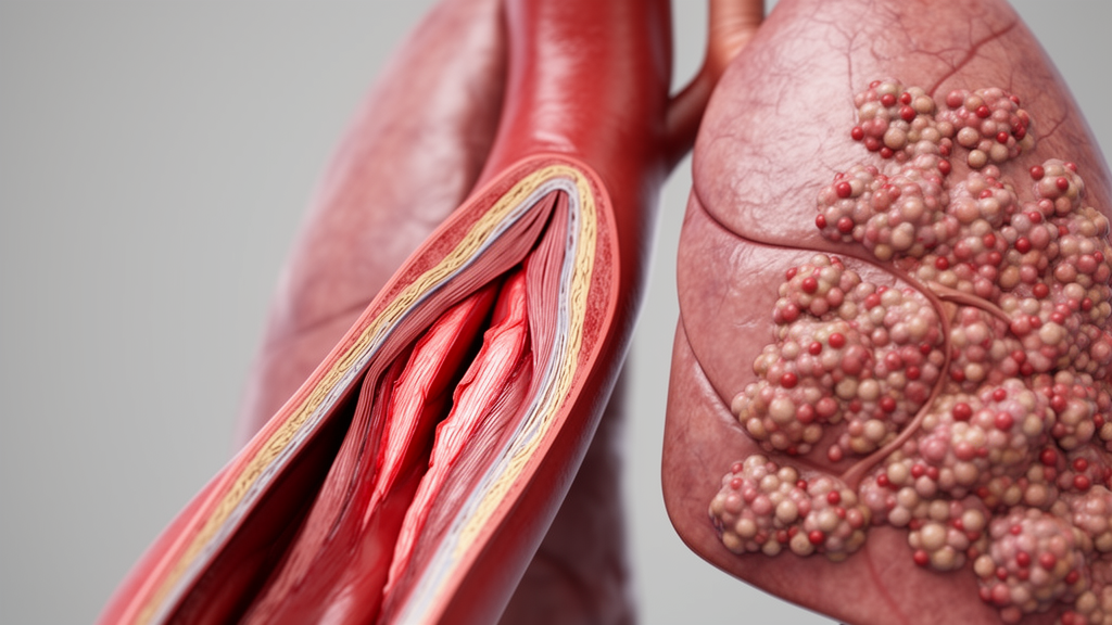
A newly published clinical investigation in thoracic oncology documents the emergence of an occult Stanford type B aortic dissection during postoperative follow-up in a patient treated for locally advanced small-cell lung cancer. The case involved a fifty-six-year-old female patient who underwent neoadjuvant chemoimmunotherapy followed by thoracic surgical resection, presenting a severe structural vascular complication despite demonstrating an exceptional tumor response and showing no typical aortic dissection symptoms such as chest or back pain. Surgery for small-cell lung cancer has conventionally been reserved for carefully staged early-stage cT1 to cT2 nodal-negative presentations, whereas concurrent platinum and etoposide chemoradiotherapy remains the established primary standard of care for more advanced limited-stage disease. While neoadjuvant chemoimmunotherapy protocols utilizing programmed death-ligand 1 inhibitors such as adebrelimab represent an active area of clinical investigation, the physiological interplay between systemic immunotherapeutic agents, cytotoxic regimens, and invasive thoracic operative techniques remains a subject requiring rigorous observation.
The patient was initially evaluated after an asymptomatic pulmonary mass in the left upper lobe was incidentally detected during a routine chest computed tomography scan in April 2026. Clinical history revealed occasional non-productive cough without episodes of hemoptysis, chest pain, or unintended weight loss. The patient had never smoked cigarettes and possessed no documented personal history of cardiovascular disorders, hypertension, or relevant familial vascular disease. Positron-emission tomography combined with computed tomography conducted in May 2026 revealed intense hypermetabolic uptake within the primary pulmonary lesion and left hilar lymph nodes, registering maximum standardized uptake values of 14.42 and 9.87 respectively. Notably, the initial baseline nuclear imaging demonstrated no pathological fluorodeoxyglucose uptake along the thoracic aortic wall. Subsequent contrast-enhanced computed tomography confirmed a lobulated mass measuring thirty by twenty-five millimeters exhibiting radiologically assessed direct invasion into the main trunk of the left pulmonary artery, which established a clinical stage of cT4N1M0 stage IIIA under the ninth edition tumor-node-metastasis staging guidelines.
Histopathological confirmation of small-cell lung cancer was established via computed tomography-guided percutaneous biopsy in late May 2026. Immunohistochemical staining confirmed that the tumor cells stained strongly positive for AE1/AE3, thyroid transcription factor 1, synaptophysin, and cluster of differentiation 56, accompanied by focal immunoreactivity for chromogranin A and a proliferation index of approximately eighty percent according to Ki-67 scoring. Baseline physical examination demonstrated stable hemodynamics, with an initial arterial blood pressure of 125/75 millimeters of mercury and a resting pulse rate of 57 beats per minute consistent with sinus bradycardia. Baseline pulmonary function testing showed preserved ventilatory capacity, with a forced expiratory volume in one second at 88.1 percent of predicted value and maximal voluntary ventilation at 90.6 percent of predicted value. Routine hematologic indices, blood chemistry panels, and baseline coagulation parameters fell entirely within normal physiological reference ranges prior to the initiation of systemic antineoplastic therapy.
Because upfront surgical resection was deemed oncologically unsuitable due to central vascular invasion and left pulmonary artery encasement, the multidisciplinary oncology team initiated neoadjuvant systemic therapy planned for two therapeutic cycles before definitive local consolidation reassessment. The regimen combined the investigational anti-PD-L1 monoclonal antibody adebrelimab at a fixed intravenous dose of 1200 milligrams with etoposide administered at 140 milligrams on days one through three and cisplatin administered at 50 milligrams on days one and two. The two planned treatment cycles commenced on May 30 and June 21, 2026, respectively. Throughout the entire neoadjuvant treatment period, the patient experienced only mild nausea and manageable vomiting, without developing severe myelosuppression, cytopenias, or other high-grade toxicities. Consequently, the clinical team did not administer recombinant granulocyte colony-stimulating factor or long-acting formulations such as pegfilgrastim at any point during the neoadjuvant chemoimmunotherapy course.
Restaging chest computed tomography performed on July 11, 2026, revealed dramatic anatomical regression of the primary parenchymal lesion and marked clearance of the associated hilar soft tissue. Preoperative contrast-enhanced brain magnetic resonance imaging excluded metastatic intracranial spread, identifying only two small, stable lesions considered likely benign meningiomas. On July 29, 2026, exactly thirty-eight days following the start of the second chemoimmunotherapy cycle, the patient underwent video-assisted thoracoscopic left upper lobectomy at an external tertiary medical center. During the minimally invasive procedure, surgeons encountered moderate hilar fibrosis and indistinct anatomical dissection planes encompassing the pulmonary hilum and subcarinal station 7 lymph nodes. Operative notes documented the utilization of an ultrasonic scalpel to achieve hemostatic control of persistent localized oozing occurring posterior to the subcarinal nodal bed in immediate proximity to the descending thoracic aorta, with no explicit mechanical wall perforation recorded.
Subsequent external histopathologic examination of the surgical specimen confirmed an outstanding therapeutic response, documenting residual primary tumor tissue measuring seventeen by ten by nine millimeters consistent with a major pathological response. Microscopic assessment verified complete microscopic resection with negative bronchial, vascular, and soft tissue margins, alongside an absence of pleural, perineural, or lymphovascular invasion. Comprehensive pathological staging evaluated twenty-four dissected regional lymph nodes harvested across stations 4L, 5, 6, 7, 8, 9, 10, 11, 12, and 13, all of which were entirely histologically negative for metastatic carcinoma. This pathological clearance yielded a downgraded post-treatment anatomical stage of ypT1N0M0. The residual neoplastic cells retained characteristic neuroendocrine marker profiles, and the patient recovered without immediate postoperative complications, remaining asymptomatic through hospital discharge following the completion of the pulmonary resection.
On August 25, 2026, the patient was admitted to the oncology service for routine clinical and diagnostic reassessment prior to scheduled adjuvant chemotherapy. Physical evaluation revealed stable vital signs, showing a systemic blood pressure of 113/72 millimeters of mercury and a regular pulse rate of 82 beats per minute, with a well-healing thoracic incision scar and an otherwise unremarkable cardiopulmonary examination. However, routine restaging contrast-enhanced computed tomography completed on August 26, twenty-eight days after the thoracic lobectomy, unexpectedly revealed structural findings indicative of a new-onset Stanford type B aortic dissection. The patient consistently denied experiencing sudden-onset tearing back pain, chest discomfort, shortness of breath, diaphoresis, or any peripheral neurological deficits. The structural aortic breakdown was completely occult from a symptomatic standpoint, having developed insidiously without subjective clinical warnings or overt hemodynamic destabilization during the postoperative recovery window.
A rigorous retrospective multi-reader radiological review of serial thoracic imaging examinations was conducted by three senior cardiovascular radiologists to clarify the chronologic timeline of the vascular injury. The baseline contrast scan from May 18, the preoperative restaging scan from July 11, and the postoperative scan from August 26 demonstrated remarkably stable mid-descending thoracic aortic dimensions at a matched ninth thoracic vertebral level, measuring 23 by 21 millimeters, 23 by 20 millimeters, and 23 by 21 millimeters, respectively. None of the sequential cross-sectional scans exhibited radiographic evidence of circumferential mural thickening, soft-tissue cuffing, or periaortic inflammatory stranding. However, multiplanar sagittal and axial reformations on the August 26 scan clearly identified a defined intimal flap separating an anterior false lumen from a posterior true lumen, alongside a focal structural discontinuity of the external aortic adventitia situated directly adjacent to the prior station 7 lymphadenectomy bed.
Given the acute structural compromise of the aortic wall and the presence of adventitial discontinuity conferring a severe hazard of free thoracic rupture, the clinical team executed urgent thoracic endovascular aortic repair on August 28, 2026. Diagnostic digital subtraction angiography localized the primary intimal tear along the lesser curvature of the thoracic aortic arch distal to the origin of the left subclavian artery, positioned in close proximity to, yet anatomically distinguishable from, the focal adventitial disruption. Combined angiographic and tomographic assessments indicated that the false lumen extended inferiorly toward the level of the eleventh and twelfth thoracic vertebrae, though the precise craniocaudal boundaries could not be definitively measured. Deployed endovascular stent graft coverage successfully excluded the entry tear and obliterated false lumen perfusion while preserving flow through the left subclavian artery, leading to an uneventful recovery and hospital discharge three days later.
Following successful aortic stabilization, the oncology service resumed multimodal cancer management on September 13, 2026, administering adjuvant adebrelimab at 1200 milligrams, followed by adjuvant etoposide and cisplatin chemotherapy beginning on September 14. Follow-up contrast-enhanced computed tomography of the chest, abdomen, and pelvis performed on October 10, forty-three days after endovascular repair, verified persistent patent lumen reconstruction through the intact stent graft without localized endoleak, alongside noncalcified plaques scattered along the aorta. In evaluating the underlying pathophysiological mechanisms, clinicians considered multiple interacting variables, including immune-mediated large-vessel vasculitis, cytotoxic endothelial toxicity, and localized perioperative mechanical trauma. Published medical literature has documented rare autoimmune aortitis and secondary aortic dissections following immune checkpoint inhibitor therapy, typically manifesting after months of therapeutic exposure and presenting with systemic symptoms, fluorodeoxyglucose avidity, or distinct circumferential periaortic wall thickening on axial tomographic imaging.
In the documented case, however, the complete absence of aortic wall thickening, the normal baseline vascular metabolic activity, and the relatively modest serum C-reactive protein measurement of 1.5 milligrams per liter preoperatively argue against diffuse immunotherapeutic aortitis as a solitary trigger. Instead, the authors highlight the close anatomical proximity between the localized adventitial breach and the surgical bed where ultrasonic energy was applied to achieve subcarinal hemostasis. The extensive hilar fibrosis and altered tissue consistency induced by neoadjuvant chemoimmunotherapy may increase the susceptibility of regional perivascular tissue planes to mechanical or thermal shear stress during minimally invasive nodal clearance. Although intraoperative vessel injury was not formally recognized at the time of resection, the forty-six-day window between the normal preoperative scan and the diagnostic postoperative scan encompasses both the operation and the postoperative recovery phase.
The authors outline several critical methodological and clinical limitations inherent to single-patient observational reports. Histopathological examination of the affected aortic wall could not be obtained because endovascular repair does not permit direct tissue sampling, leaving microscopic vascular inflammatory status unconfirmed. Additionally, intermittent non-invasive blood pressure recordings spanning 96 to 125 millimeters of mercury systolic cannot exclude occult transient hypertensive spikes during recovery, meaning an unexpected spontaneous dissection cannot be completely ruled out. Furthermore, short-term tomographic follow-up demonstrating patent stent reconstruction cannot establish the definitive long-term vascular safety of resuming platinum chemotherapy and immune checkpoint inhibition after aortic repair. Nevertheless, this notable clinical experience underscores that life-threatening aortic pathology can evolve silently during multimodal cancer treatment, emphasizing the necessity of meticulous serial aortic evaluation on routine oncologic imaging.
Subject of Research: New-onset asymptomatic type B aortic dissection following multimodal chemoimmunotherapy and surgical resection for small-cell lung cancer
Article Title: Case Report: New-onset type B aortic dissection after chemoimmunotherapy and lung resection for small-cell lung cancer
Article References: Lu, F., Liu, J., Zhang, Y., Deng, L., Ma, H., & Feng, W. (2026). Case Report: New-onset type B aortic dissection after chemoimmunotherapy and lung resection for small-cell lung cancer. Frontiers in Oncology, 16, Article 2024707. https://doi.org/10.3389/fonc.2026.2024707
Image Credits: AI Generated
DOI: 10.3389/fonc.2026.2024707
Keywords: aortic dissection, type B aortic dissection, small-cell lung cancer, chemoimmunotherapy, adebrelimab, cisplatin, etoposide, thoracic endovascular aortic repair, video-assisted thoracoscopic surgery, lobectomy, computed tomography, ultrasonic scalpel, intimal flap, adventitial discontinuity, lymphadenectomy, vascular complication, thoracic oncology
News Source: Nathaniel Bowman. (November 19, 2026). Symptomless Type B Aortic Dissection Detected After Small-Cell Lung Cancer Resection. Scienmag.