In the relentless pursuit of advancing breast cancer diagnostics, a groundbreaking study published in BMC Cancer introduces superb microvascular imaging (SMI) as a transformative tool for evaluating axillary lymph nodes (ALNs). This innovative imaging technique offers a remarkable leap forward in distinguishing benign from malignant lymph nodes, significantly enhancing pre-surgical diagnostics. The investigative team embarked on a prospective clinical study, enrolling over a hundred patients presenting with suspicious axillary lymph node findings, thus laying the groundwork for robust, clinically meaningful insights into the vascular architecture of these lymph nodes.
Axillary lymph nodes serve as pivotal anatomical structures in breast cancer staging and management, often dictating the therapeutic pathway and prognosis. Traditional ultrasound methodologies, while valuable, have faced limitations in accurately characterizing lymph node vascular supply. The advent of SMI addresses these challenges by enabling refined visualization of microvascular patterns without the drawbacks of contrast agents or invasive procedures. By capturing both semi-quantitative features, such as the number and distribution of vascular branches, and qualitative characteristics including the appearance of these vessels, researchers have gained unprecedented clarity in discerning pathological processes.
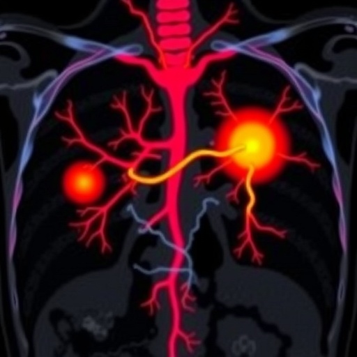
The technical prowess of SMI lies in its advanced Doppler algorithm, which isolates and depicts low-velocity blood flow within microvascular networks, overcoming noise interference seen in conventional power Doppler ultrasound (PDUS). This enhanced sensitivity facilitates a detailed evaluation of subtle vascular anomalies that correlate strongly with malignant transformation. In the study, 108 lymph nodes from 102 patients were meticulously analyzed, with post-imaging confirmation achieved through histopathology or rigorous clinical follow-up, thereby ensuring diagnostic accuracy and reliability in the findings.
A central aspect of the investigation was the diagnostic performance comparison of various vascular feature combinations observed through SMI. The synergy between distribution and appearance of vessels emerged as the most potent predictor of malignancy, achieving an area under the curve (AUC) of 0.776 with near 79% accuracy in receiver operating characteristic (ROC) analyses. This dual-parameter evaluation surpasses the capabilities of singular vascular descriptors alone and underscores the value of integrating multidimensional imaging data for clinical decision-making.
Moreover, the study illuminated the robustness of SMI through interobserver agreement assessments conducted by seasoned radiologists with varying experience levels. The substantial concordance observed in interpreting SMI images exempts A from concerns regarding operator dependency, assuring reproducibility and clinical applicability across different healthcare settings. This facet is especially critical when transitioning novel imaging technologies from research domains into widespread diagnostic practice.
Notably, the diagnostic prowess of SMI was accentuated among patients with confirmed primary breast cancer. Here, the accuracy soared to an impressive 92.76%, coupled with an elevated AUC of 0.855, signaling the method’s exceptional capability to correctly stratify lymph node pathology in this high-risk subgroup. This level of precision drastically benefits clinicians by minimizing unnecessary invasive procedures, thereby sparing patients undue morbidity while optimizing treatment planning.
SMI’s non-invasive nature and superior visualization not only elevate diagnostic confidence but also promise enhanced patient comfort and streamlined clinical workflows. Unlike contrast-enhanced imaging methods, SMI obviates the risks and costs associated with contrast administration, making it a patient-friendly alternative readily integrable into routine ultrasound examinations. This technological advancement aligns with contemporary trends emphasizing precision medicine and tailored therapeutic interventions.
Underlying the success of SMI is its ability to elucidate the intricate microvascular networks within lymph nodes, revealing heterogeneity indicative of pathological neovascularization. Malignant nodes characteristically exhibit chaotic, irregular vascular patterns, in stark contrast to the organized vasculature of benign nodes. By capturing these nuanced differences, SMI operates as a sensitive biomarker for tumor-induced angiogenesis, offering insights that extend beyond morphology into functional tissue characterization.
This pioneering research sets a precedent for future multicenter trials and integration of SMI with other imaging modalities such as elastography and contrast-enhanced ultrasound. Combining these complementary technologies could herald a new epoch in non-invasive lymph node assessment, further refining diagnostic algorithms and potentially enabling real-time intraoperative evaluations. The capacity to confidently differentiate lymph node status preoperatively has significant ramifications for surgical planning and patient prognosis.
The implications of adopting SMI in clinical breast cancer pathways are transformative. Enhanced diagnostic accuracy leads to better risk stratification, individualized treatment regimens, and potentially improved survival rates. By reducing false negatives and false positives, healthcare systems can allocate resources more effectively, decreasing the burden of overtreatment or delayed intervention. These systemic benefits reinforce the imperative to embrace cutting-edge imaging innovations that augment clinical precision.
Beyond breast cancer, the application of superb microvascular imaging extends to myriad pathological conditions where microvascular remodeling is a hallmark, including inflammatory diseases, vascular anomalies, and other malignancies. The successful demonstration of SMI in axillary lymph node evaluation thus paves the way for broader adoption across diverse medical disciplines, fostering interdisciplinary advances in diagnostic imaging.
The meticulous methodology employed in this research, featuring blinded independent reviews by experts and the largest cohort of suspicious ALNs studied using SMI to date, consolidates its scientific rigor. Such comprehensive analyses diminish biases and enhance the reproducibility of conclusions, thereby strengthening the evidence base supporting SMI as a next-generation diagnostic modality. This study embodies the confluence of technological innovation and clinical acumen driving progress in oncology diagnostics.
In summary, the adoption of superb microvascular imaging marks a significant milestone in breast cancer evaluation, enabling precise differentiation of suspicious axillary lymph nodes through detailed vascular characterization. The fusion of semi-quantitative and qualitative assessments propels diagnostic accuracy to unprecedented levels, fostering improved clinical outcomes while streamlining patient care pathways. As this technology continues to be validated and refined, it holds promise to revolutionize imaging protocols, exemplifying the future of non-invasive cancer diagnostics.
The research team’s contribution heralds a new era where vascular imaging details are integrated seamlessly into routine practice, empowering clinicians with actionable insights. By transcending the limitations of conventional ultrasound modalities, SMI stands poised to become a cornerstone technique in breast cancer diagnosis and beyond. Ongoing innovation and clinical evaluation will undoubtedly expand its capabilities, ultimately benefiting patients worldwide through earlier detection and personalized treatment strategies.
Subject of Research: Diagnostic evaluation of axillary lymph nodes in primary breast cancer using superb microvascular imaging (SMI).
Article Title: Diagnostic value of super microvascular imaging in differentiating axillary lymph nodes: semi-quantitative and qualitative approach in primary breast cancer and suspicious axillary nodes.
Article References:
Tokur, O., Aydin, S., Kilinc, F. et al. Diagnostic value of super microvascular imaging in differentiating axillary lymph nodes: semi-quantitative and qualitative approach in primary breast cancer and suspicious axillary nodes. BMC Cancer 25, 1705 (2025). https://doi.org/10.1186/s12885-025-14936-w
Image Credits: Scienmag.com
DOI: 10.1186/s12885-025-14936-w (Published 04 November 2025)
Tags: advanced Doppler algorithm in imagingaxillary lymph node diagnosisbreast cancer diagnosticsclinical study on lymph nodesdistinguishing benign and malignant lymph nodesenhancing cancer staging through imagingimaging techniques for lymph nodesmicrovascular patterns visualizationnon-invasive lymph node evaluationpre-surgical diagnostics innovationsuperb microvascular imagingvascular architecture in breast cancer


