Credit: Niigata University
Niigata, Japan- New insights into the three-dimensional (3D) morphology of the human uterine endometrium could advance our understanding of the mechanisms of endometrial regeneration and fertilized egg implantation while clarifying the pathogenesis of menstrual disorders, infertility and endometrium-related diseases such as adenomyosis, endometriosis, endometrial hyperplasia and endometrial cancer.
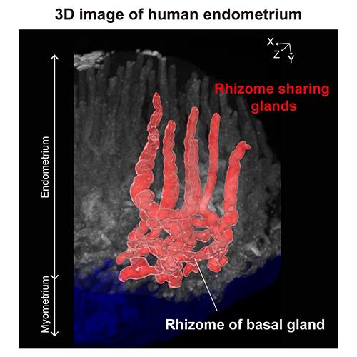
The endometrial glands are comprised of complicated winding and branching structures, and conventional 2D imaging techniques have been unable to adequately assess their shape. This limitation has prevented elucidation of the mechanisms of endometrial regeneration during the menstrual cycle and the location of endometrial progenitor cells. Recent developments in 3D tissue-clearing imaging allowed researchers from Niigata University to explore the endometrial structure in greater depth.
The team, led by Prof. Takayuki Enomoto, Dr. Kosuke Yoshihara and Dr. Manako Yamaguchi of the Department of Obstetrics and Gynecology, Graduate School of Medical and Dental Sciences, Niigata University, implemented an updated clear, unobstructed brain/body imaging cocktail and computational analysis (CUBIC) protocol with fluorescent microscopy to obtain 3D full thickness images of the human uterine endometrium. As expected, the 3D tissue analysis revealed unique morphologies that had not been previously detected by 2D histological observation. Human endometrial glands were discovered to form a complex plexus network in the stratum basalis referred to as the ‘rhizome’ structure. Named after the rhizomatous plant’s stems, the rhizome describes the horizontally expanding plexus morphology of the basal glands. Furthermore, some glands share the rhizome and rise toward the luminal epithelium. Interestingly, these structural features were detected in all samples, regardless of age or menstrual cycle phase, which suggests they are basic components of the normal human endometrium.
After observing the normal structure of the uterine endometrium, the researchers expanded their analysis to clarify the 3D structure of adenomyotic lesions. Adenomyosis is a benign condition characterized by the atypical presence of endometrial glands and stroma within the myometrium, or the muscular walls of the uterus. Prior to this study, various hypotheses regarding the etiology of adenomyosis existed, including endometrial invasion, endometriotic invasion and de novo metaplasia. Reconstituted 3D images verified the endometrium breaches the myometrium, where these aberrant structures lengthen to form fine glands that intricately branch and extend along the large blood vessels of the uterus, analogous to an ant colony.
The human endometrium is a dynamic tissue that exerts morphological and functional changes on a monthly basis in response to ovarian hormones. This highly regenerative tissue is involved in menstruation and implantation of the fertilized egg, giving it a central role in women’s reproductive health. Unfortunately, the regenerative nature of the endometrial glands can encourage the development and progression of ‘endometrium-related diseases’, such as adenomyosis, endometriosis, endometrial hyperplasia and endometrial cancer. The pathogenesis of endometrium-related conditions remains unclear, which has hindered the development of effective preventative measures and therapeutic strategies.
In their previous 2018 genomic study, the investigators sought to provide insight into the origins of endometriosis and elucidate associated molecular characteristics. They found that the genes most recurrently mutated in endometriosis-associated ovarian cancers were also frequently mutated in endometriotic epithelium, and, surprisingly, in normal uterine endometrial glands. These novel findings led to the hypothesis that clonal genomic alternations may change the structure of endometrial glands and increase susceptibility to endometrium-related diseases. Establishing the 3D morphology of normal uterine endometrium was therefore essential to testing this model and ultimately sparked this investigation.
As pointed out by principal investigator Prof. Enomoto “Our 3D imaging established the baseline for the 3D structure of human endometrial glands and adenomyotic lesions. These findings dynamically change the concept of human uterine endometrium.”
While extensive research in the field of obstetrics and gynecology is required to identify the mechanisms of normal endometrium function and related diseases, this study can provide novel insight into the 2D shape of the human endometrial glands for the first time in nearly one hundred years along with the contemporary 3D model of the tissue. The potential application of this work is not lost on Prof. Enomoto “The 3D representation of the human endometrium will lead to a better understanding of the human endometrium in various fields, including histology, pathology, pathophysiology, reproduction and oncology.”
###
Media Contact
Takayuki Enomoto
[email protected]
Original Source
https:/
Related Journal Article
http://dx.


