The zero-calorie sweetener stevia has long been marketed as a guilt-free alternative to sugar, but a new study suggests the plant may do far more than simply sweeten coffee. Researchers in Iran report that an aqueous extract of Stevia rebaudiana appears to protect the livers of diabetic rats by suppressing ferroptosis, an iron-dependent form of cell death that has emerged as a key player in diabetes-related organ damage. The findings, published in Molecular Biology Reports, add a surprising molecular dimension to a sweetener already known for its glucose-lowering and antioxidant properties.
Ferroptosis is unlike the better-known forms of programmed cell death such as apoptosis. Instead of the tidy cellular dismantling seen in apoptosis, ferroptosis is a violent, oxidative demise: iron catalyzes the peroxidation of lipids in cell membranes, causing them to rupture. Because liver tissue is rich in iron-handling machinery and constantly exposed to metabolic stress, it is particularly vulnerable. In diabetes, chronically elevated blood sugar floods cells with reactive oxygen species, depletes antioxidant defenses, and tips the delicate redox balance toward lipid peroxidation, making ferroptosis a plausible driver of diabetic liver injury.
To test whether stevia could intervene in this process, the team led by Zahra Amirahmadi of Islamic Azad University, Kazerun, together with colleagues at Shiraz University of Medical Sciences, induced diabetes in rats using streptozotocin, a chemical that destroys insulin-producing pancreatic beta cells. Twenty-four animals were randomly divided into four groups of six: healthy controls receiving water, diabetic rats receiving water, diabetic rats given an aqueous stevia extract at 400 milligrams per kilogram of body weight, and diabetic rats treated with metformin at 500 milligrams per kilogram, the standard first-line diabetes drug. After 28 days of daily dosing, the researchers measured blood chemistry, antioxidant capacity, inflammatory markers, the expression of ferroptosis-related genes, cell death by TUNEL assay, and liver tissue architecture.
The biochemical results were striking. Compared with untreated diabetic animals, stevia-treated rats showed significantly reduced fasting blood sugar, with the authors reporting a p-value below 0.01. The liver enzymes alanine aminotransferase and aspartate aminotransferase, both classic markers of hepatocyte damage, dropped substantially, indicating that the extract was protecting liver cells from injury. Body weight, which typically falls in uncontrolled diabetes, partially recovered in the stevia group. Meanwhile, the animals’ antioxidant defenses rebounded: total antioxidant capacity rose, and serum levels of the enzymes glutathione peroxidase, superoxide dismutase, and catalase all increased significantly, each with a p-value below 0.001.
Inflammation, another hallmark of diabetic liver disease, also receded. Levels of the inflammatory signaling molecules interleukin-6, C-reactive protein, tumor necrosis factor alpha, interleukin-1 beta, and the transcription factor NF-kappa-B were all lower in stevia-treated rats than in diabetic controls. This matters because inflammation and ferroptosis feed each other in a vicious cycle: dying cells release damage signals that recruit immune cells, which in turn produce reactive oxygen species that push neighboring cells toward ferroptosis. Breaking that loop at either end can blunt the entire cascade.
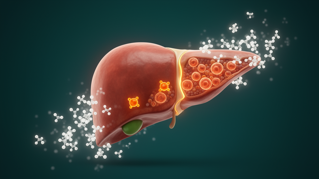
The most novel part of the study lies in the gene expression data. Stevia treatment upregulated GPX4, the glutathione peroxidase enzyme that sits at the very center of ferroptosis control by converting dangerous lipid peroxides into harmless lipid alcohols. It also boosted FTH1, the ferritin heavy chain that sequesters free iron and thereby removes the catalyst ferroptosis depends on, and SLC7, part of the system xc- cystine importer that supplies cells with the building blocks for glutathione, GPX4’s essential cofactor. All three genes are canonical suppressors of ferroptosis, and their coordinated upregulation suggests stevia acts on the pathway at multiple points rather than through a single target.
Histology and cell death assays corroborated the molecular story. TUNEL staining, which labels cells with fragmented DNA, revealed significantly fewer dying cells in the livers of stevia-treated diabetic rats, and microscopic examination showed improved tissue morphology compared with the disorganized, damaged architecture of untreated diabetic livers. Malondialdehyde, a breakdown product of lipid peroxidation and a direct chemical fingerprint of ferroptotic damage, was significantly reduced. Taken together, the data sketch a coherent mechanism: stevia restores antioxidant capacity, sequesters iron, repairs the glutathione supply line, and thereby prevents the membrane lipid destruction that defines ferroptosis.
The study is not without caveats. It involved only 24 rats over 28 days, and the streptozotocin model produces a form of insulin-deficient diabetes that does not fully recapitulate the more common type 2 disease in humans. The authors measured gene expression rather than protein levels or enzyme activity for the ferroptosis regulators, and they did not use a ferroptosis-specific inhibitor to prove that the pathway is causally responsible for the observed protection. Dose translation is another open question: 400 milligrams per kilogram in a rat is a large intake relative to what a human would consume in sweetened beverages, and purified steviol glycosides used in commercial sweeteners differ chemically from whole aqueous extract.
Even so, the work fits into a growing body of evidence linking plant-derived compounds to ferroptosis control. Previous studies have shown that polyphenols such as kaempferol, naringenin, and curcumin can suppress ferroptosis through the Nrf2 antioxidant pathway, and earlier work by the same Shiraz group found that stevia extract alleviates endoplasmic reticulum stress in diabetic rat livers. Other research has demonstrated that synthetic steviol derivatives protect against cardiomyopathy by inhibiting ferroptosis, and that stevia can prevent experimental liver cirrhosis by modulating profibrotic pathways. The new study extends this literature by explicitly connecting stevia to the ferroptosis machinery in diabetic liver injury.
For now, the practical takeaway is cautious. Stevia remains a safe, calorie-free sweetener with an expanding portfolio of documented biological activities, and this study strengthens the case that its polyphenol-rich extract carries genuine pharmacological potential. But rat data at high doses do not justify treating diabetes-related liver disease with sweetener packets, and human clinical trials would be needed before any therapeutic claims could be made. What the research does offer is a mechanistic roadmap: if ferroptosis is a driver of diabetic complications, then identifying safe dietary compounds that switch the pathway off, as this stevia extract appears to do in rats, could open a new front in the fight against the metabolic disease epidemic.
Subject of Research: Protective effects of aqueous Stevia extract against ferroptosis-mediated liver damage in streptozotocin-induced diabetic rats
Article Title: Regulating ferroptosis: the impact of stevia on alleviating liver damage in streptozotocin-induced diabetic rats
Article References: Amirahmadi, Z., Raeisi, A., Koohpeyma, F., Moghadam, D., Kiani, R., Jamshidi, S., Shams, F., Naeimi, S., & Dastghaib, S. (2026). Regulating ferroptosis: the impact of stevia on alleviating liver damage in streptozotocin-induced diabetic rats. Molecular Biology Reports, 53(1), Article 1686. https://doi.org/10.1007/s11033-026-12804-9
Image Credits: AI Generated
DOI: 10.1007/s11033-026-12804-9
Keywords: stevia, ferroptosis, diabetes, liver injury, streptozotocin, GPX4, oxidative stress, antioxidants, inflammation, ferritin, lipid peroxidation, metformin
News Source: Daisy Hatcher. (October 9, 2026). Stevia Extract Shields Diabetic Livers by Taming Iron-Driven Cell Death. Scienmag.