For the first time in the realm of developmental biology, a groundbreaking stem cell model has astonishingly generated a structure analogous to the human yolk sac during early embryogenesis, originating from a single stem cell population without any genetic manipulation. This unprecedented achievement, realized by researchers at the University of Michigan Engineering, ushers in a novel era for studying human development outside the constraints of natural embryos and offers promising pathways toward understanding conditions like early pregnancy loss.
In traditional embryology, the yolk sac is understood to emerge from hypoblast cells—distinct from the epiblast lineage that gives rise to the primary embryo. Remarkably, the Michigan team’s model—comprised solely of epiblast-like pluripotent stem cells—spontaneously formed yolk-sac-like structures. This defies canonical developmental dogma by revealing previously unappreciated plasticity in epiblast cells, suggesting that they possess latent capabilities to differentiate beyond embryo proper tissues when guided by precise environmental cues.
This breakthrough builds on the team’s established expertise in applying mechanical signals as developmental architects. Unlike genetic engineering approaches that have artificially directed stem cells toward yolk sac fates, Jianping Fu’s team implemented geometric confinement techniques to spatially pattern the stem cell population. By confining human pluripotent stem cells into a micropatterned, 0.8-millimeter diameter disc—mirroring the size of the epiblast disc during gastrulation—they created a controlled physical microenvironment conducive to natural self-organization and differentiation.
To initiate gastrulation-like processes, the team supplemented the culture medium with bone morphogenetic protein 4 (BMP-4), a critical developmental morphogen normally secreted by extra-embryonic tissues in vivo. BMP-4 signaling, in concert with intrinsic cell-cell interactions under geometric constraint, triggered the stem cells to differentiate into distinct lineages that organized into a trilaminar embryonic disc-like configuration. This multi-layered structure paralleled the germ layers of the nascent embryo, including ectodermal, mesodermal, and endodermal analogs.
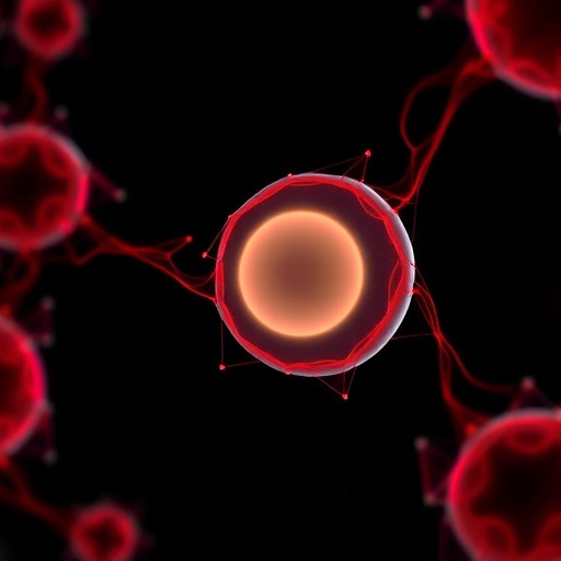
Unexpectedly, in addition to the three germ layers, the model generated an amniotic sac-like cavity lined with amnion cells on the external side. Most notably, on the internal side, a yolk-sac-like structure emerged, characterized by a cavity enveloped by endodermal-like cells. This morphology aligns vividly with the architecture of the early human yolk sac, critical for nutrition and the initiation of hematopoiesis before placental circulation is established.
Confirming the identity of these yolk sac analogs was a pivotal challenge. The researchers utilized lineage tracing techniques, introducing a fluorescent reporter driven by gastrulation-specific gene activation to chart the differentiation trajectory of the epiblast cells. The presence of the transcription factor HNF4A, a hallmark of yolk sac and early visceral endoderm development, further substantiated the stem cells’ differentiation into yolk sac lineage. The team collaborated with colleagues at the Chinese Academy of Sciences, who provided post-implantation monkey embryo data to cross-validate the molecular signatures observed.
It is critical to underscore that this level of embryo-like development, reaching features between 16 to 21 days post-fertilization, transcends the widely observed 14-day culture limit for human embryonic models. However, since this system is transgene-free and stem cell–based rather than derived from actual embryos, it bypasses many ethical and technical barriers, enabling extended observation of developmental phenomena that were previously inaccessible.
While these models displayed remarkable fidelity in recapitulating early human embryogenesis, they do not perfectly mirror all developmental facets. For instance, the absence of trophoblast cells—which form the placenta—and an abnormal thickness of the three-layered embryonic disc indicate limitations in the model’s biological completeness. Furthermore, after initial organization, the cultures tend to lose structural integrity and deviate from embryo-like features, suggesting that additional biochemical and mechanical cues are necessary for sustained morphogenesis.
The stem cell models were meticulously crafted using micropatterned culture plates produced at the University of Michigan Lurie Nanofabrication Facility, combining cutting-edge bioengineering with molecular biology tools. The researchers’ comprehensive analysis relied on advanced microscopy, genomics, histology, and flow cytometry, leveraging core facilities across the university to validate cellular identity, gene expression dynamics, and tissue architecture. This multidisciplinary approach exemplifies the convergence of engineering and biology in modern developmental science.
This discovery carries significant implications beyond basic science. Early pregnancy loss remains a perplexing clinical challenge, with a majority of spontaneous miscarriages occurring within the first few weeks after fertilization. By elucidating the mechanisms governing yolk sac formation and early embryonic patterning, such stem cell models could illuminate the causes of developmental arrest or defects, potentially informing diagnostic and therapeutic strategies to improve reproductive health outcomes.
Moreover, the use of mechanical confinement and biochemical gradients to guide pluripotent stem cell self-organization paves the way for refined tissue engineering techniques. These principles might be extended to model other human developmental stages and diseases, thereby facilitating drug discovery, toxicology screening, and regenerative medicine applications. Efforts to commercialize this technology are underway, with patent applications and partnerships being sought to translate these models from bench to bedside.
In a broader context, this work challenges longstanding paradigms about embryonic lineage specification and cell fate plasticity. The capacity of epiblast-like stem cells to generate extra-embryonic structures without genetic reprogramming underscores the power of the microenvironment and biophysical cues in directing cell behavior. Such insights elevate our understanding of human biology’s foundational processes and highlight the potential of stem cell engineering to recreate complex developmental systems in vitro.
Looking ahead, refining these models to incorporate missing cell types like trophoblasts, improving structural stability, and extending developmental timelines will be critical milestones. Integration with emerging technologies like single-cell multi-omics, live imaging, and computational modeling may unlock deeper mechanistic insights. As these embryo-like constructs become increasingly sophisticated, they promise to reshape our capacity to interrogate human development ethically and with unprecedented resolution.
To conclude, the University of Michigan’s achievement in engineering a pluripotent stem cell model that autonomously generates yolk-sac-like structures represents a transformative leap in developmental biology. By marrying mechanical engineering principles with cellular biology, this work provides a robust platform for investigating early human embryogenesis, potentially revolutionizing reproductive medicine and tissue engineering.
Subject of Research: Human embryonic development, stem cell differentiation, and yolk sac formation
Article Title: A transgene-free, human peri-gastrulation embryo model presents trilaminar embryonic disc-, amnion- and yolk sac-like structures
News Publication Date: 2024
Web References:
Nature Cell Biology Article DOI: 10.1038/s41556-026-01930-y
University of Michigan Mechanical Engineering Faculty – Jianping Fu
Lurie Nanofabrication Facility
References:
Fu, J. et al. “A transgene-free, human peri-gastrulation embryo model presents trilaminar embryonic disc-, amnion- and yolk sac-like structures.” Nature Cell Biology (2024).
Keywords: human embryogenesis, pluripotent stem cells, yolk sac, gastrulation, mechanical confinement, BMP-4 signaling, epiblast, stem cell differentiation, amniotic sac, developmental biology, tissue engineering, early pregnancy loss
Tags: early embryogenesis in vitroepiblast cell plasticitygeometric confinement of stem cellshuman developmental biology modelsmechanical signaling in stem cell patterningmicropatterned stem cell culturemodeling early pregnancy lossnon-genetic manipulation in stem cell researchnovel approaches to human embryo developmentpluripotent stem cells differentiationstem cell model of human embryoyolk sac generation without hypoblasts

