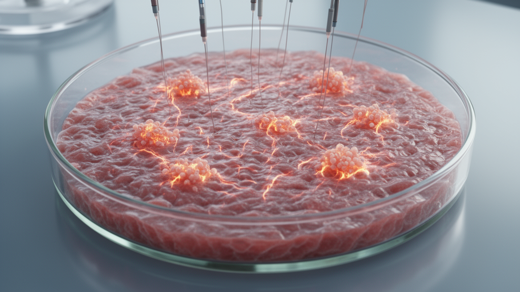
Human stem-cell cultures can be pushed toward an atrial fibrillation-like state by combining heart muscle cells derived from induced pluripotent stem cells with fibroblasts and subjecting them to repeated rapid electrical stimulation, according to a laboratory study published in Frontiers in Cardiovascular Medicine. The two-dimensional model reproduced several measured features associated with atrial fibrillation, or AF: abnormal electrical activity, disturbed calcium handling, slow or blocked conduction, fibrotic structural change and molecular signals linked to stress and fibrosis. The findings establish a platform for investigating disease mechanisms and screening potential therapies, but the researchers emphasize that it reproduces selected local features rather than the full biology of a human heart.
The platform was built around human induced pluripotent stem cell-derived atrial cardiomyocytes, or hiPSC-aCMs, which were differentiated, purified and matured on an extracellular matrix. The researchers first compared these atrial cells with ventricular cardiomyocytes produced through the same general stem-cell workflow. Atrial cells had a higher frequency of spontaneous activation, a shorter calcium-transient duration and a higher oxygen-consumption rate. They also expressed more atrial natriuretic peptide, myosin light chain 7 and the potassium-channel gene KCNA5, while KCNJ3 expression was higher but not statistically significant. Although the transcription factor NR2F2 was less abundant in the atrial cultures, the authors said its expression can change during maturation and that the combined functional and molecular profile supported atrial identity. Maturation was associated with increased cardiac troponin I, phospholamban and the calcium-release channel RYR2.
To recreate the electrical stress associated with rapid rhythms, the investigators developed an intermittent tachypacing protocol. Each hour, cultures received 45 minutes of stimulation at 1.5 Hz followed by 15 minutes at 5 Hz, with the sequence repeated for seven days. The electrical field was applied at 20 V/cm with 0.5-millisecond pulses. In cardiomyocyte-only cultures, this intervention increased the frequency of spontaneous action potentials and shortened action-potential duration at 80% repolarization. It also increased action-potential triangulation, while leaving the upstroke slope unchanged, and was associated with faster action-potential conduction. The authors described these changes as consistent with a tachycardia-like electrical phenotype, while noting that the stimulation frequencies were regimen settings rather than confirmed cellular activation frequencies: one-to-one capture was not verified throughout treatment, and continuous optical monitoring was not performed during the seven-day protocol.
Optical mapping also revealed changes in intracellular calcium cycling after intermittent tachypacing. The variability in spontaneous calcium-release frequency rose by 332% compared with untreated cultures. Calcium-transient upstroke became slower, calcium-release amplitude declined and calcium waves propagated more slowly across the monolayer. Beat-to-beat variability in calcium reuptake also increased, although the duration of calcium reuptake did not significantly change and basal diastolic calcium remained similar. Following a burst-pacing arrhythmia inducibility test, treated cells had a reduced calcium upstroke slope and amplitude, together with diminished restoration of diastolic calcium. These results show that intermittent tachypacing disturbed both the regularity and strength of calcium cycling in the atrial cell cultures.
The second component of the model was co-culture with human adult atrial cardiac fibroblasts, which supplied a fibrotic environment. Researchers tested mixtures containing 100%, 90% or 70% hiPSC-aCMs, with the balance made up by fibroblasts. The strongest structural remodeling occurred in the 70:30 cultures. Collagen III expression increased by more than 100-fold, and the cytokine TGFB1 was also elevated. The co-culture produced a 61% reduction in excitability, a 130% increase in action-potential duration at 80% repolarization and a 143% increase in triangulation. Action-potential upstroke fell by 66%, while conduction velocity decreased and conduction block occurred in a measurable proportion of the field. Calcium handling also deteriorated: the calcium upstroke slope declined by 50%, calcium-transient amplitude fell by 34%, and spontaneous calcium-release frequency became more variable. These changes grew with fibroblast abundance, showing that the ratio between the two cell populations affected the phenotype.
When intermittent tachypacing and fibroblast co-culture were combined, the model displayed an AF-like mix of increased cellular activity, shortened repolarization and impaired conduction. Treated 70:30 cultures showed a 25% increase in spontaneous activity, a 16% shortening of action-potential duration at 80% repolarization and a 27% reduction in conduction velocity relative to paced cardiomyocyte-only cultures. The comparison was important because adding fibroblasts did not further accelerate spontaneous activation, but it substantially impeded the electrical wave. The authors therefore proposed that fibroblasts may contribute primarily to conduction slowing and structural heterogeneity, while the pacing protocol supplies the tachycardia-associated electrical remodeling.
Molecular measurements offered possible explanations for the functional findings. Western blot analysis showed that total phospholamban, abbreviated PLN, was reduced after intermittent tachypacing in both cardiomyocyte-only and 70:30 co-cultures. Phosphorylated PLN and the calcium pump SERCA2a did not change significantly, but the ratio indicating the balance between PLN regulation and SERCA2a was elevated in paced 70:30 cultures. The investigators interpreted this shift as evidence of disturbed calcium reuptake capacity, although they also noted that PLN messenger RNA increased, suggesting that transcript abundance did not straightforwardly predict the amount of functional protein. RYR2 and ATP2A2 messenger RNA levels were unchanged. By contrast, the stress- and remodeling-related genes NPPA, NPPB and TGFB1 increased, with the largest increases in the paced fibroblast co-culture. The molecular pattern linked the functional changes to altered calcium regulation and pro-fibrotic signaling, although the experiments did not establish a single causal pathway for every electrical abnormality.
Several limitations constrain what the platform can represent. It contains atrial cardiomyocytes and fibroblasts but omits macrophages, endothelial cells, smooth-muscle cells and sympathetic and parasympathetic neurons, so it captures local remodeling cues without systemic regulation. The final proportions of cells in the co-cultures were estimated from immunofluorescence rather than confirmed by flow cytometry. The experiments also did not incorporate patient-specific genetic predispositions to AF, and optical mapping measured the combined behavior of a cellular network rather than resolving ion currents in individual cardiomyocytes. The study lacked a time-matched control for continuous 1.5-Hz stimulation, preventing the investigators from separating the effects of the two pacing frequencies. Caffeine-induced calcium-release assays were also not used to determine the relative contribution of sarcoplasmic-reticulum calcium to calcium decay, leaving that mechanism incompletely resolved.
Even with those caveats, the results show that the model can reproduce interacting AF-associated changes in a controlled human-cell system. The fibroblast module supplied a fibrotic, conduction-limiting substrate; intermittent tachypacing added stress-associated electrical remodeling; and the molecular analysis linked both to altered calcium cycling and pro-fibrotic signaling. The authors say the platform could be expanded to examine additional AF triggers, including sympathetic and parasympathetic influences, and potentially integrated into established proarrhythmia-safety frameworks such as CiPA and JiCSA. It may also support mechanistic investigation and evaluation of therapeutic interventions. As an in vitro modeling study, however, the work did not test patient benefit, clinical treatment effectiveness or whether compounds screened in the system predict clinical performance.
Subject of Research: A human two-dimensional stem-cell model of atrial fibrillation-associated electrical and fibrotic remodeling
Article Title: hiPSC-derived new approach methodology (NAM) recapitulates hallmarks of atrial fibrillation
Article References: Kozlowski, P., Muthukumarasamy, K. M., Buranello, T. W., Jo, S., Deshmukh, A., Monteiro da Rocha, A., & Oral, H. (2026). hiPSC-derived new approach methodology (NAM) recapitulates hallmarks of atrial fibrillation. Frontiers in Cardiovascular Medicine, 13, Article 1814075. https://doi.org/10.3389/fcvm.2026.1814075
Image Credits: AI Generated
DOI: 10.3389/fcvm.2026.1814075
Keywords: atrial fibrillation, induced pluripotent stem cells, atrial cardiomyocytes, cardiac fibroblasts, intermittent tachypacing, calcium handling, cardiac fibrosis, optical mapping, electrophysiology, new approach methodologies, hiPSC-derived, approach
News Source: Ophelia Keating. (October 20, 2026). Stem-cell cultures reproduce selected atrial fibrillation features in vitro. Scienmag.