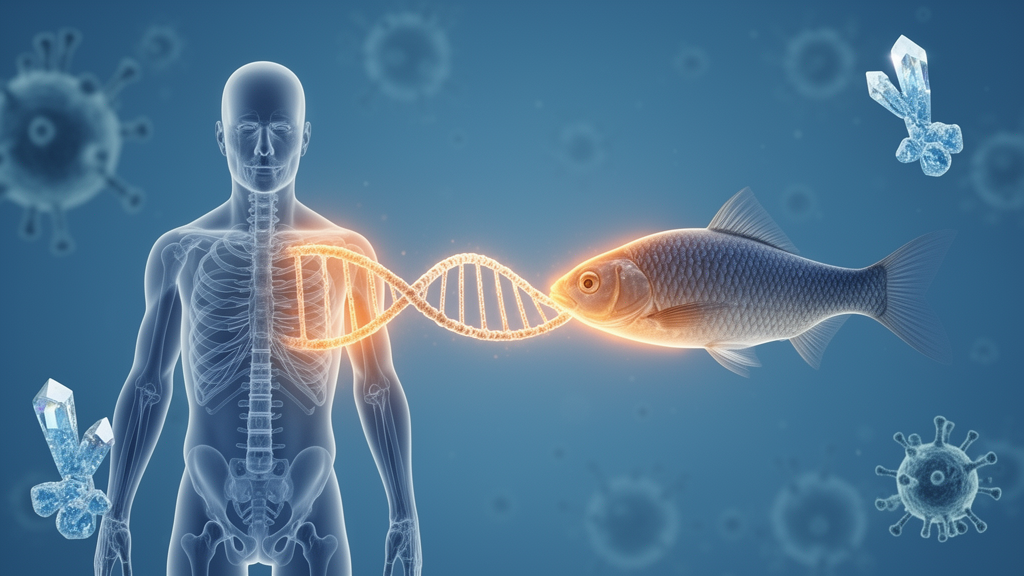
More than forty years ago, researchers fishing for the secrets of calcium balance in teleost fish isolated a glycoprotein from a small endocrine organ called the corpuscles of Stannius. The protein, later named stanniocalcin, acted as a hypocalcemic hormone, lowering serum calcium when levels climbed too high. Because those organs exist only in bony fish and holosteans, scientists assumed stanniocalcin was a specialized adaptation to aquatic life. Then came a surprise: a homolog was discovered in human tissues, and a second human paralog soon followed, leading to the designations STC1 and STC2. A new open-access review in Molecular Biology Reports by Krista Agathe Bossow, Caroline Marie Norup Kjeldsen, and Claus Oxvig of Aarhus University now takes stock of what these evolutionarily ancient proteins actually do in mammals, and the answer is far stranger than the original fish story suggested.
Structurally, both mammalian stanniocalcins are secreted, glycosylated, covalent homodimers, with monomeric units of 247 and 302 amino acids respectively. Phylogenetic analyses place their homologs in early vertebrate evolution, and predicted STC-like sequences appear as far back as single-celled eukaryotes, hinting at deep evolutionary roots and fundamental cellular functions. Yet for decades their mammalian roles remained murky. Knockout mice lacking Stc1 showed no anatomical or histological abnormalities and, remarkably, maintained normal serum calcium and phosphate even after vitamin D3-induced hypercalcemia. Double knockouts lacking both genes were equally normal in mineral homeostasis, suggesting the stanniocalcins do not play a major role in mineral balance in mice. The most striking phenotype instead appeared in growth: mice lacking Stc2, and double knockouts, grew roughly fifteen percent faster and larger than wild-type littermates after weaning.
The mechanistic explanation arrived only about ten years ago, and it now constitutes the best-founded molecular function of both proteins. STC1 and STC2 potently inhibit the pappalysin metalloproteinases PAPP-A and PAPP-A2. These enzymes promote insulin-like growth factor signaling by cleaving IGF binding proteins, a family of six carriers that hold IGF-1 and IGF-2 in a bioinactive state. PAPP-A cuts IGFBP-2, -4, and -5, while PAPP-A2 cleaves IGFBP-3 and -5. When the pappalysins release IGF from these restraints, the freed growth factor activates the IGF receptor and downstream PI3K/Akt pathways, driving cellular proliferation, differentiation, and organismal growth. By blocking the pappalysins, the stanniocalcins keep IGF locked away, reducing bioactive signaling and growth.
Recent cryo-electron microscopy structures have clarified exactly how this works. Two independent research groups solved the structure of the 2:2 PAPP-A:STC2 heterotetramer, revealing that STC2 adopts a homodimeric, all-helical fold positioned at the upper central exosite of the PAPP-A dimer, sterically blocking IGFBP substrate entry without touching the active site. Crucially, cysteine 120 of STC2 forms a disulfide bond with cysteine 732 of PAPP-A, making the inhibition covalent and irreversible. STC1 lacks an equivalent cysteine and instead inhibits through high-affinity non-covalent interactions, with a dissociation constant of 75 picomolar against PAPP-A. Both stanniocalcins also inhibit PAPP-A2 in vitro, though far less is known about that interaction. The genetic evidence converges beautifully: mice overexpressing either STC1 or STC2 exhibit severe postnatal dwarfism with a 30 to 45 percent size reduction, comparable to the 40 percent dwarfism of Pappa knockout mice. In humans, children carrying unstable or inactive PAPP-A2 variants show decreased growth, and a large genome-wide association study identified an STC2 variant, Arg44Leu, that increases carrier height by 2.1 centimeters and impairs PAPP-A complex formation. Mice expressing a STC2 Cys120Ala mutant unable to form the covalent bond grow normally, confirming the mechanism.
Beyond the IGF system, the review critically evaluates a decade of reports implicating the stanniocalcins in calcium handling, immune modulation, mitochondrial stress responses, integrin signaling, DNA repair, Notch signaling, and reproduction. The evidence here is generally less robust, often resting on co-immunoprecipitation experiments of varying control quality, and the reported effects are frequently context-dependent. This is particularly true for STC1, which can exert opposing effects depending on cell type and biological setting. Proposed intracellular and nuclear activities also raise unresolved questions about how a secreted glycoprotein is taken up and trafficked within cells.
In calcium homeostasis, the picture is gradually coming into focus through the IGF axis. In zebrafish, STC1 suppresses epithelial calcium uptake by regulating ionocyte quiescence and proliferation in an IGF-dependent manner, and recent work shows the STC1-PAPP-A-IGF pathway helps maintain acoustic startle thresholds in larvae, with cadherin 16 acting as a negative regulator of STC1. STC2, meanwhile, has been linked to suppression of store-operated calcium entry, possibly through interaction with the ER calcium sensor STIM1, as shown by co-immunoprecipitation in several cell types and human platelets. But the exact mechanisms remain unclear, and the review emphasizes the need to disentangle how calcium regulation depends on the stanniocalcins’ established roles in IGF signaling.
Immune modulation is one of the most actively explored areas. One of the best-supported roles of STC1 involves calreticulin, an ER protein that functions as an eat-me signal when presented on the cell surface. Across murine tumor models and human cancer cell lines, STC1 traffics calreticulin from the plasma membrane to the mitochondria, reducing phagocytosis by macrophages and dampening T cell activation, a mechanism described as a phagocytosis checkpoint driving tumor immune resistance. The anti-inflammatory protein A20 stabilizes STC1 by inhibiting GSK3β-mediated degradation, reinforcing this pathway. STC1 also engages βPIX to release and activate the transcription factor YAP, promoting M2 macrophage polarization, while separate reports show STC1 sequestering GRP94 to inhibit macrophage maturation, or binding the IGF2 receptor to reduce interleukin-1β secretion. STC2 contributes through complex formation with histidine-rich glycoprotein, quantified at a dissociation constant of 55 nanomolar, which suppresses macrophage phagocytosis and maturation. Depending on context, the stanniocalcins can be pro- or anti-inflammatory, reflecting their complexity in immune microenvironments.
Mitochondrial regulation and oxidative stress represent some of the most cited but most intricate functions. Secreted STC1 is internalized through megalin, a membrane receptor whose signal peptide leucines are crucial for the interaction, and trafficked to mitochondria via Rab32-dependent vesicular transport, where it co-immunoprecipitates with Sirtuin 3 and modulates respiration, glycolysis, and redox state. STC1 activates the AMPK pathway, upregulating uncoupling proteins and SIRT3, though the direction of its effect on ATP production appears ambiguous and context-dependent. Both stanniocalcins contain a heme regulatory motif with a cysteine proposed to bind hemin, and both engage the Nrf2/HO-1/hemin antioxidant pathway. STC1 protects hemin from hydrogen peroxide-mediated decay, while STC2, but not STC1, interacts with heme oxygenase-1 through amino acids 181 to 200, potentially delivering hemin as a substrate for degradation. A recent study connected several of these threads, showing that STC1’s interaction with calreticulin activates CAMKK2, which in turn activates the AMPK-GSK3β-Nrf2-HO-1 axis, reducing oxidative stress and apoptosis. Still, the review notes that how and when STC1 activates AMPK, and whether the stanniocalcins cooperate or counteract each other in Nrf2 regulation, remain open questions.
In DNA damage repair, STC1 exhibits strikingly dual effects. On the protective side, it enhances homologous recombination-mediated repair and cell survival after irradiation by co-immunoprecipitating with BRCA1 and promoting its recruitment to damage sites. On the damaging side, under oxidative stress STC1 stabilizes PARP1 by inhibiting its ubiquitination, prolonging its activity and promoting PARP1-dependent cell death, or parthanatos, in colon-derived cells. STC1 has also been implicated as a SUMO-1 E3 ligase, increasing SUMOylation of RanGAP-1 and thereby affecting protein stability. STC2, by contrast, appears consistently protective, increasing radioresistance in esophageal cancer cells by co-immunoprecipitating with a repair complex of PRMT5, CLNS1A, and WDR77, and correlating with radioresistance in lung cancer. In reproduction, the stanniocalcins are repeatedly linked to ovarian physiology, pregnancy, and lactation, with circulating STC1 rising in maternal serum in both humans and mice, ovarian tissue containing unique high molecular weight Big STC1 variants of approximately 85, 112, and 135 kilodaltons that reduce progesterone secretion from luteal cells, and STC2 associating with RanBPM and the androgen receptor in human testis. Yet no well-founded mechanism explains these reproductive effects.
The review concludes that inhibition of PAPP-A and regulation of IGF signaling remain the only stanniocalcin functions substantiated through biomolecular structures, biochemical, genetic, and functional evidence working in concert. Everything else, from calcium handling to immunity to DNA repair, is supported to varying degrees, often through co-immunoprecipitation alone, and many findings warrant further experimental analysis to establish whether the interactions are direct, specific, and functionally relevant. Determining where, when, and how STC1 and STC2 act, and to what extent their diverse reported effects converge on shared mechanisms, will be essential for developing a coherent understanding of stanniocalcin biology and for exploiting their increasingly reported importance in human physiology and disease.
Subject of Research: Molecular Biology
Article Title: Stanniocalcin-1 and −2: The insulin-like growth factor system and beyond
Article References: Bossow, K. A., Kjeldsen, C. M. N., & Oxvig, C. (2026). Stanniocalcin-1 and −2: The insulin-like growth factor system and beyond. Molecular Biology Reports, 53(1), Article 1724. https://doi.org/10.1007/s11033-026-12940-2
Image Credits: AI Generated
DOI: 10.1007/s11033-026-12940-2
Keywords: stanniocalcin, PAPP-A, insulin-like growth factor, IGF signaling, protein interactions, cellular signaling, molecular biology reports, Stanniocalcin-1, insulin-like, growth, factor, system
News Source: Drew Townsend. (October 17, 2026). Stanniocalcins: From Fish Calcium Hormone to Master Regulator of Human Growth. Scienmag.