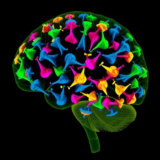
In a groundbreaking study published in Nature Neuroscience, researchers have leveraged spatial proteomic analysis to uncover the intricate microenvironment-dependent states of microglial cells within human Alzheimer’s disease (AD) brains. This pioneering work, led by Sanchez-Molina, Rosmus, Brownell, and colleagues, offers profound insights into the cellular complexity underlying neurodegenerative pathology, potentially setting the stage for targeted therapeutic interventions.
Alzheimer’s disease represents an ever-growing global health crisis characterized by progressive cognitive decline and widespread neurodegeneration. Despite decades of research, the precise molecular and cellular dynamics contributing to its pathogenesis remain elusive. Microglia, the resident immune cells of the central nervous system, have emerged as pivotal players, implicated both in neuroprotective roles and in exacerbation of pathology via neuroinflammation. However, the heterogeneity of microglial states and their contextual relationships with the surrounding brain microenvironment has posed significant challenges to detailed characterization.
The study harnesses advanced spatial proteomics, a cutting-edge approach combining high-dimensional protein profiling with spatial localization, to map protein expression directly within intact human brain tissue sections. By applying multiplexed immunohistochemistry and mass spectrometry imaging, the investigators achieved an unprecedented resolution of microglial phenotypes and their spatial distribution relative to AD pathological hallmarks such as amyloid plaques and neurofibrillary tangles.
One of the core revelations from this research is the identification of distinct microglial cell states that differ fundamentally depending on their microenvironment. Microglia situated proximal to amyloid plaques displayed activated and pro-inflammatory proteomic signatures, whereas those in plaque-distant regions exhibited homeostatic or even neuroprotective profiles. This spatial heterogeneity underscores microglia’s adaptive capacity and suggests that local cues within the brain milieu drive their functional polarization.
Significantly, the proteomic data revealed novel markers and pathways differentiating these microglial subpopulations. Proteins involved in phagocytosis, complement cascade activation, and cytokine signaling were enriched near plaques, implicating an immune response skewed toward clearance and inflammation. Conversely, microglia in unaffected areas were characterized by proteins supporting tissue maintenance and synaptic modulation, hinting at a role in preserving neuronal networks despite widespread pathology.
The methodological rigor of the study is notable. Tissue samples from postmortem human brains diagnosed with AD were carefully processed to preserve spatial integrity. Subsequent imaging and protein quantification allowed the team to not only catalogue protein expression profiles but also to spatially map these molecular signatures relative to neuropathological landmarks within the same tissue. This dual capability is transformative, bridging molecular biology and histopathology in a way that traditional bulk tissue analyses cannot match.
Importantly, the findings challenge the prevailing notion of microglia as a monolithic population within the diseased brain. Instead, they advocate for a model in which microglial cells assume diverse, context-dependent states governed by precise molecular programs influenced by their immediate surroundings. Such heterogeneity likely contributes in complex ways to disease progression, balancing neuroprotective responses and neurotoxic outcomes.
Another compelling aspect of the study is its potential translational impact. Understanding the proteomic landscape and spatial context of microglial states opens avenues for precision targeting of dysfunctional microglia in AD. Therapeutic strategies could aim to modulate the harmful microglial subsets engaged in chronic inflammation while preserving or enhancing those involved in tissue repair and homeostasis.
Furthermore, this work offers a valuable resource for biomarker discovery. Proteins uniquely expressed or enriched in specific microglial states could serve as molecular signatures accessible through cerebrospinal fluid or imaging techniques, enhancing diagnostic accuracy, disease staging, and monitoring of therapeutic responses in clinical settings.
The study also highlights the broader applicability of spatial proteomics to other neurological disorders. Diseases such as Parkinson’s, multiple sclerosis, and traumatic brain injury feature complex neuroimmune interactions that might similarly be unraveled by this approach. Thus, this research not only advances the understanding of AD but also establishes a versatile platform for neurodegenerative research as a whole.
Critically, the integration of spatial proteomics with other omics technologies promises to deepen insights into AD etiology. Correlating proteomic profiles with transcriptomics, metabolomics, and epigenomic data, all within spatial frameworks, will enrich the multidimensional characterization of disease microenvironments.
The authors also emphasize the need for longitudinal studies to track the evolution of microglial states over disease progression. Such temporal analyses could illuminate when specific microglial phenotypes emerge and how they influence or respond to neurodegeneration and pathology accumulation.
In addition to revealing molecular underpinnings, the study contributes fundamental knowledge at the intersection of neuroimmunology and spatial biology. It expands the conceptual framework of brain function and dysfunction, recognizing that cellular identity and activity cannot be fully understood in isolation from spatial context.
While this research is a monumental step forward, challenges remain. The technical complexity, demand for high-quality human tissue samples, and computational analysis hurdles highlight the necessity for interdisciplinary collaboration and technological innovation to translate these findings into clinical practice.
Given the scale and scope of Alzheimer’s disease worldwide, approaches that uncover such detailed mechanistic insights are urgently needed. This study exemplifies how cutting-edge technologies can illuminate previously hidden aspects of brain pathology, driving forward the quest for effective interventions.
As the research community continues to embrace spatially resolved proteomics and allied methods, a new era of neurodegenerative disease research dawns. The ability to decode cellular heterogeneity in situ promises not only to clarify pathophysiology but also to inspire the design of smarter, localized therapeutic strategies.
In conclusion, the spatial proteomic analysis conducted by Sanchez-Molina and colleagues represents a transformative advance in Alzheimer’s disease research, revealing microenvironment-dependent microglial cell states with diverse functional implications. Their findings challenge existing dogma, enrich our molecular understanding of AD, and open the door for innovative diagnostic and therapeutic approaches that could reshape treatment paradigms in neurodegeneration.
Subject of Research:
Spatial proteomic characterization of microglial cell states in human Alzheimer’s disease brain tissue.
Article Title:
Spatial proteomic analysis in human Alzheimer’s disease brains enables identification of microenvironment-dependent microglial cell states.
Article References:
Sanchez-Molina, P., Rosmus, DD., Brownell, D. et al. Spatial proteomic analysis in human Alzheimer’s disease brains enables identification of microenvironment-dependent microglial cell states. Nat Neurosci (2026). https://doi.org/10.1038/s41593-026-02267-3
Image Credits:
AI Generated
DOI:
https://doi.org/10.1038/s41593-026-02267-3
Tags: advanced proteomic techniques for neurodegenerative diseasescellular complexity in Alzheimer’s pathologymass spectrometry imaging in neurosciencemicroenvironment-dependent microglial statesmicroglia and amyloid plaque interactionmicroglial cell heterogeneity in neurodegenerationmultiplexed immunohistochemistry for brain tissueneurofibrillary tangles spatial analysisneuroinflammation and microglial rolesprotein expression mapping in brain tissuespatial proteomics in Alzheimer’s diseasetargeted therapeutic strategies for Alzheimer’s


