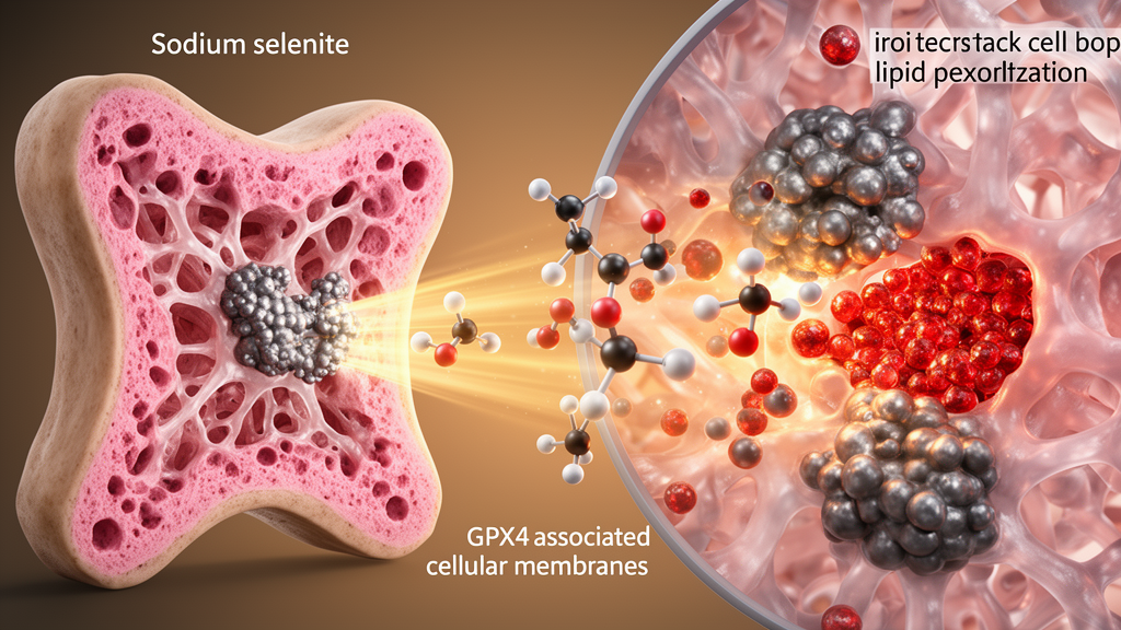
Sodium selenite reduced oxidative damage and restored bone-forming capacity in mouse bone marrow stromal cells exposed to hydrogen peroxide, according to a study combining cell experiments, RNA sequencing, gene-silencing tests and an ovariectomized mouse model of osteoporosis. The treatment also limited bone loss in the animals, with the higher of two tested doses producing the greater improvement in trabecular structure. Molecular analyses identified glutathione peroxidase 4 (GPX4), a selenium-dependent antioxidant enzyme, as a central mediator: sodium selenite increased its thermal stability, and silencing GPX4 removed much of the compound’s ability to suppress lipid peroxidation or promote osteogenic differentiation. These findings provide a mechanistic preclinical rationale for studying sodium selenite as a ferroptosis-directed strategy for bone regeneration, but the experiments do not establish an effective or safe human osteoporosis treatment.
The research focused on osteoblasts and their precursor bone marrow stromal cells, which normally help form bone and can be damaged by oxidative stress. Osteoporosis involves disturbed bone remodeling and deterioration of bone microarchitecture, and excessive lipid peroxidation in osteoblasts can impair their function and contribute to further skeletal deterioration. Ferroptosis is a regulated form of cell death associated with iron-dependent lipid peroxidation. GPX4 limits this process through the GPX4–SLC7A11–glutathione antioxidant axis, but the study asked whether sodium selenite could protect bone-forming cells by engaging that system. Researchers used third-passage rat bone marrow stromal cells and selected 0.1 micromolar sodium selenite for the principal in vitro experiments after preliminary cytotoxicity screening and physiological selenium-tolerance assessments.
In the first cell experiments, hydrogen peroxide markedly impaired the osteogenic program. It suppressed the osteogenic proteins RUNX2, SP7 and COL1A1, reduced alkaline phosphatase activity, and limited mineralization of the extracellular matrix. Sodium selenite counteracted these effects in a dose-dependent manner: it increased alkaline phosphatase staining, promoted formation of mineralized nodules, and raised the cellular signals associated with RUNX2 and osteocalcin. Cell viability was not significantly affected by 0.1 micromolar sodium selenite, indicating that the observed changes were not attributed to fewer viable cells. These measurements assessed early osteogenic commitment, matrix mineralization and selected protein markers, rather than demonstrating that the treatment produced mature bone in culture.
To investigate why the cells were protected, the investigators performed RNA sequencing after 72 hours of hydrogen peroxide exposure with or without 0.1 micromolar sodium selenite. The comparison identified 329 upregulated and 336 downregulated genes. Separate KEGG and Gene Ontology analyses converged on ferroptosis-related functions, including iron transport and homeostasis, while gene set enrichment analysis indicated suppression of a ferroptosis pathway signature in the sodium-selenite-treated cells. These results implicated ferroptosis in the experimental response, but transcriptomic enrichment alone could not establish that pathway as the cause of improved osteogenesis. The researchers therefore compared sodium selenite with ferrostatin-1, a pharmacological ferroptosis inhibitor, and directly measured several cellular features associated with the process.
Both treatments produced similar patterns in the cell-based assays. Hydrogen peroxide increased intracellular reactive oxygen species, malondialdehyde, a byproduct of lipid peroxidation, and the labile ferrous iron detected by FerroOrange. It also depleted glutathione. Sodium selenite reduced reactive oxygen species and malondialdehyde, restored glutathione, and largely eliminated the excess labile iron signal. Analysis of mitochondrial membrane potential with JC-1 and measurements of total intracellular iron also changed in the same protective direction. Western blotting and quantitative reverse-transcription PCR showed treatment-associated changes in GPX4, SLC7A11 and COX2, proteins linked to ferroptosis control. The parallel response to ferrostatin-1 supported the interpretation that sodium selenite acts as an inhibitor of ferroptosis, although the comparison was conducted in cultured cells under an acute oxidative-stress model rather than in people with osteoporosis.
GPX4 was then examined as a possible direct target. Molecular docking predicted that sodium selenite could fit complementarily within a predicted GPX4 binding pocket, but a docking result alone does not demonstrate physical binding. The researchers therefore used a cellular thermal shift assay, which indicated that sodium selenite increased the melting temperature of GPX4 from 43.44°C to 47.25°C, a shift of 3.81°C consistent with stabilization through direct interaction. Small interfering RNA was then used to reduce GPX4 expression. GPX4 silencing lowered the osteogenic proteins RUNX2, SP7 and COL1A1 and also reduced SLC7A11. Sodium selenite could no longer significantly recover these osteogenic markers after GPX4 knockdown, and its capacity to restore alkaline phosphatase activity, mineralization, glutathione levels and low oxidative stress was substantially diminished. The knockout-by-silencing experiments therefore support GPX4 as necessary for much of the protective effect observed in this cellular system.
The investigators next tested the treatment in female C57BL/6 mice. Thirty animals were randomly assigned to four groups of five: sham-operated controls, ovariectomized controls, and ovariectomized mice receiving daily intraperitoneal sodium selenite at 0.05 or 0.1 milligrams per kilogram. Injections began one week after surgery and continued for 14 consecutive days, with assessment eight weeks after surgery. Ovariectomy produced substantial trabecular bone loss. Micro-computed tomography and three-dimensional reconstruction showed that sodium selenite increased trabecular number and improved bone morphometric measures, with more pronounced changes at 0.1 milligrams per kilogram. The treatment also reduced trabecular separation and porosity, increased the volume of newly formed bone, and improved the organization of trabecular structure. Calcein double labelling indicated increased mineral deposition, consistent with greater new bone formation during the experimental interval.
The study also examined whether the selected doses produced detectable organ toxicity. Histological examination of the heart, lungs and kidneys showed no apparent morphological abnormalities in the sodium-selenite-treated animals. Serum alanine aminotransferase, aspartate aminotransferase, blood urea nitrogen and creatinine did not show pathological elevations compared with the sham and ovariectomized groups. These findings provide limited evidence of biosafety over the short treatment and observation period used in the study; they cannot establish the safety margin for human use or rule out adverse effects during chronic exposure. Similarly, the mouse results demonstrate preservation and formation of bone in an ovariectomy model, not fracture prevention in patients. The groups were also small, with five animals per group, and the experiment did not include an established osteoporosis drug as a comparator.
Several limitations constrain the broader interpretation of the work. The in vitro model used an acute hydrogen peroxide insult, which cannot reproduce the multiple biological and mechanical causes of osteoporosis. The animal experiment was relatively short, included only young adult female mice after ovariectomy, and did not establish whether the skeletal benefits persist after treatment stops. Mechanistic experiments concentrated on osteogenic differentiation, leaving unresolved whether sodium selenite also directly affects osteoclast-mediated bone resorption. The authors proposed that selenium may support GPX4 through several routes, including stabilization of the protein, provision of selenium for selenocysteine incorporation, and possible activation of the Nrf2–Keap1 pathway, but those specific mechanisms were not established in the reported experiments. Longer studies in additional osteoporosis models, together with direct assessment of resorption and extended safety monitoring, will be needed before translational conclusions can be drawn.
Overall, the experiments connect sodium selenite, GPX4 and ferroptosis in a coherent preclinical chain. The compound protected cultured bone-forming cells from hydrogen peroxide-associated oxidative damage, enhanced osteogenic differentiation, and showed a GPX4-dependent effect on lipid peroxidation, glutathione and iron homeostasis. In ovariectomized mice, the same treatment was associated with preservation of trabecular architecture, increased new bone formation and no obvious short-term hepatic or renal abnormalities in the tested dose range. The work is best viewed as a mechanistic study of a candidate bone-protective strategy rather than evidence that selenium supplementation can treat human osteoporosis. Its central implication is that GPX4-mediated restraint of ferroptosis may be a useful target for future interventions aimed at protecting osteoblasts, subject to confirmation in broader, longer and clinically relevant models.
Subject of Research: Preclinical investigation of sodium selenite, GPX4-mediated ferroptosis inhibition and bone protection in ovariectomized mice
Article Title: GPX4‐Mediated Inhibition of Ferroptosis by Sodium Selenite Ameliorates Bone Metabolic Disorders in Ovariectomized Mice
Article References: Luo, W., Xu, H., Liu, X., Zhang, Y., Zhou, H., Pan, X., Geng, D., & He, H. (2026). GPX4 ‐Mediated Inhibition of Ferroptosis by Sodium Selenite Ameliorates Bone Metabolic Disorders in Ovariectomized Mice. Journal of Cellular and Molecular Medicine, 30(20), Article e71414. https://doi.org/10.1111/jcmm.71414
Image Credits: AI Generated
DOI: 10.1111/jcmm.71414
Keywords: sodium selenite, ferroptosis, GPX4, osteoporosis, bone loss, osteogenesis, oxidative stress, lipid peroxidation, bone marrow stromal cells, ovariectomy, Mediated, Inhibition
News Source: Drew Townsend. (October 21, 2026). Sodium selenite curbs bone loss in mice through GPX4-linked ferroptosis inhibition. Scienmag.