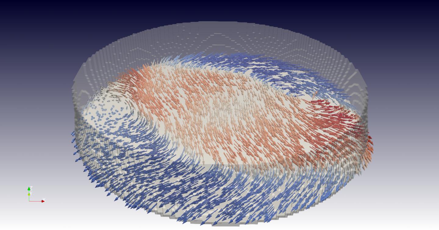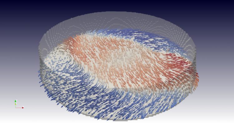
Credit: Paul Scherrer Institute/Claire Donnelly
For the first time, researchers at the Paul Scherrer Institute PSI have recorded a “3D film” of magnetic processes on the nanometer scale. This reveals a variety of dynamics inside the material, including the motion of swirling boundaries between different magnetic domains. The insights were gained with a method newly developed at the Swiss Light Source SLS. It could help to make magnetic data storage devices more compact and efficient. The researchers are publishing the results of their investigations today in the journal Nature Nanotechnology.
A magnetic sticker staying attached to a refrigerator door is hardly surprising. However, when one approaches the nanometre range (where one nanometer is one millionth of a millimeter), physicists still find magnets, and their behaviour, puzzling. At the same time, the effects occurring on this small scale are highly relevant for future technologies. Now, for the first time, PSI researchers were able to record a short “film” of the three-dimensional magnetic structure inside a material with nanoscale resolution.
“Magnetism plays a role in many ways in our everyday lives; but at this very small, fundamental level, the phenomena are not yet fully understood”, explains Claire Donnelly, lead author of the study. Donnelly was a researcher at PSI at the time of the experiment and now works at the University of Cambridge in the UK.
The researchers used X-ray light from the Swiss Light Source SLS at PSI and a special tomographic method they recently developed there, which they call “time-resolved ptychographic laminography”. Their team consisted of scientists at PSI and ETH Zurich, as well as in the UK. The sample they examined consisted of a gadolinium-cobalt compound patterned into a circular disc.
More than four days for seven images
“With our method we can non-destructively scan the material and from the data reconstruct several successive 3D images of the inner magnetic structure”, says PSI researcher Manuel Guizar-Sicairos. “We can visualise the orientation of the magnetic moment at every measured point in the material and represent them as tiny magnetic compass needles.”
Just like magnetic filings, these compass needles react to an external magnetic field and to each other, forming intricate patterns throughout the entire object. The patterns contain areas – so-called domains – in which the magnetisation points predominantly in one direction. The transitions between two such areas, i.e., the domain walls, are of particular interest to researchers: “People have proposed using them as memory bits, which could possibly be used to pack data even more tightly than when using the domains”, says Donnelly. The details of these domain walls have only recently been made visible in 3D at PSI, among other places, using state-of-the-art imaging methods.
In the present study, the researchers went one step further by mapping the motion of both the domains and the domain boundaries. “We have taken seven snapshots showing points in time that are only a quarter of a billionth of a second apart. In these we can see how a domain boundary moves back and forth.” It took the scientists a little more than four days of constant measuring to collect the data, which later yielded this sequence of seven images.
Like stroboscopic light
The observed movement of the domain boundary was repeatedly and specifically induced by the researchers themselves through an externally applied magnetic field. Their images were therefore not actually recorded within a quarter of a billionth of a second. Instead, the scientists created a time loop of the changing magnetic field and took images at different points in time within it- similar to stroboscopic light seemingly slowing down a repetitive movement.
The recording of the 3D images from inside the sample in turn draws on a basic principle from computed tomography (CT). Similar to medical CT scans, the X-rays were used to take many radioscopic images of the sample one after the other, each from a slightly different angle. From the data collected, the researchers were able to recover their 3D maps of the magnetisation using software they had developed for this purpose.
“With this method, we have not just achieved time-resolved 3D movies of the interior of an object”, says Donnelly happily. “We also have been able to map the nanoscale dynamics in a magnet. In other words, we have shown that our new technique is really relevant to the development of new technology.” And Guizar-Sicairos adds: “Our new method is also suitable for other materials and could therefore have many more useful applications in the future.”
###
Text: Paul Scherrer Institute/Laura Hennemann
Images are available for download at https:/
About PSI
The Paul Scherrer Institute PSI develops, builds and operates large, complex research facilities and makes them available to the national and international research community. The institute’s own key research priorities are in the fields of matter and materials, energy and environment and human health. PSI is committed to the training of future generations. Therefore about one quarter of our staff are post-docs, post-graduates or apprentices. Altogether PSI employs 2100 people, thus being the largest research institute in Switzerland. The annual budget amounts to approximately CHF 407 million. PSI is part of the ETH Domain, with the other members being the two Swiss Federal Institutes of Technology, ETH Zurich and EPFL Lausanne, as well as Eawag (Swiss Federal Institute of Aquatic Science and Technology), Empa (Swiss Federal Laboratories for Materials Science and Technology) and WSL (Swiss Federal Institute for Forest, Snow and Landscape Research).
Additional information
Diving into magnets – Press release from 20. July 2017: http://psi.
Contact
Dr. Manuel Guizar-Sicairos
Coherent X-Ray Scattering Group
Paul Scherrer Institute, Forschungsstrasse 111, 5232 Villigen PSI, Switzerland
Telephone: +41 56 310 34 09, e-mail: [email protected] [English, Spanish]
Dr. Claire Donnelly
Cavendish Laboratory, JJ Thomson Ave, Cambridge, CB3 0HE
Telephone: +44 12 23 74 69 05, e-mail: [email protected] [English]
Original Publication
Time-resolved imaging of three-dimensional nanoscale magnetization dynamics
C. Donnelly, S. Finizio, S. Gliga, M. Holler, A. Hrabec, M. Odstrčil, S. Mayr, V. Scagnoli, L. J. Heyderman, M. Guizar-Sicairos, J. Raabe
Nature Nanotechnology 24. February 2020 (online)
DOI: 10.1038/s41565-020-0649-x
Media Contact
Dagmar Baroke
41-563-102-111
Original Source
https:/
Related Journal Article
http://dx.


