


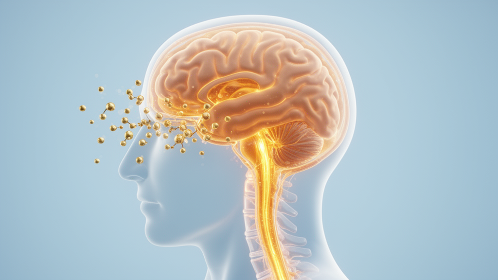
Women newly diagnosed with idiopathic intracranial hypertension (IIH) show a distinct excess of sex steroid hormones in their cerebrospinal fluid, and laboratory experiments suggest those hormones can activate the choroid plexus, the brain structure that produces cerebrospinal fluid.
IIH is a neurometabolic disorder most commonly affecting young females with obesity, and it presents with daily headaches and papilledema caused by raised intracranial pressure. Although hormonal dysfunction has long been suspected in the disease, the connection between altered steroid hormones and choroid plexus physiology remained unclear. The study’s stated objective was therefore to define the steroid hormone profile in newly diagnosed, treatment-naive patients and to identify how steroid hormones influence choroid plexus function.
The clinical arm enrolled a prospectively recruited cohort of 56 female patients with newly diagnosed, treatment-naive IIH, alongside 34 healthy controls matched for age, sex and body mass index. The researchers performed comprehensive steroid profiling on paired serum and cerebrospinal fluid samples using liquid chromatography tandem mass spectrometry, quantifying 18 steroids. In a separate subgroup, they also assessed steroid profiles in 10 patients who had entered remission, using matching samples taken at the time of diagnosis.
The profiling revealed a distinct steroid signature in patients. In cerebrospinal fluid, six steroids differed from controls, while three differed in serum. The progestins pregnenolone and progesterone, and the androgens testosterone and dehydroepiandrosterone-sulphate, were all higher in the cerebrospinal fluid of patients than controls, with pregnenolone and testosterone reaching statistical significance at q < 0.01 and progesterone at q < 0.05. In serum, progesterone was higher, while the androgens dehydroepiandrosterone and dihydrotestosterone were lower than in controls.
Notably, the deviations tracked disease activity. At remission, cerebrospinal fluid levels of pregnenolone and testosterone had normalized, suggesting that steroid dysfunction in these patients is reversible and linked to the active disease state rather than a fixed trait. The authors describe the patients as showing profound steroid hormone deviation in which steroid dysfunction normalizes at remission.
To probe what those hormonal shifts might do, the team turned to complementary preclinical experiments in rats and mice. Choroid plexus tissue was exposed to sex steroid hormones, and the researchers assessed mRNA sequencing and calcium dynamics afterwards. Both progesterone and testosterone increased intracellular calcium levels in the tissue, each with a reported significance of P < 0.0001. In the preclinical work, progesterone and testosterone each increased intracellular calcium levels (P < 0.0001), and the authors suggest that these hormones can activate the choroid plexus.
The authors are careful about what this means mechanistically. Their conclusion is that the findings suggest a potential link between steroid hormones and choroid plexus dynamics that could influence cerebrospinal fluid secretion and contribute to raised intracranial pressure, making it an interesting future study target in IIH pathophysiology. In other words, the human data establish an association between active disease and steroid excess in cerebrospinal fluid, while the animal and tissue experiments provide a plausible cellular mechanism that now requires further investigation.
Several caveats frame the work. The clinical comparison is a case-control design, which demonstrates differences between groups rather than proving that steroid excess causes raised intracranial pressure. The mechanistic activation evidence comes from rodent and cellular models, not from human choroid plexus under raised pressure. The remission subgroup was small, involving 10 patients with paired samples. The article was also shared early as a peer-reviewed, accepted manuscript carrying a permanent DOI, a version subject to further edits before the final Version of Record appears.
Even so, the study adds a concrete piece to a long-standing puzzle.
Subject of Research: Steroid hormone profiles and choroid plexus activation in idiopathic intracranial hypertension
Article Title: Cerebrospinal fluid sex steroid excess in newly diagnosed patients with idiopathic intracranial hypertension activates choroid plexus
Article References: Kamp-Jensen, C., Lylloff, T., Skovsted, M. O., Korsbaek, J. J., Hansen, N. S., Haanes, K. A., Beier, D., Hamann, S., Kogelman, L. J. A., Styrishave, B., Jensen, R. H., & Westgate, C. S. J. (2026). Cerebrospinal fluid sex steroid excess in newly diagnosed patients with idiopathic intracranial hypertension activates choroid plexus. Fluids and Barriers of the CNS. https://doi.org/10.1186/s12987-026-00880-9
Image Credits: AI Generated
DOI: 10.1186/s12987-026-00880-9
Keywords: idiopathic intracranial hypertension, cerebrospinal fluid, sex steroid hormones, choroid plexus, progesterone, testosterone, pregnenolone, intracranial pressure, case-control study, liquid chromatography mass spectrometry, neurometabolic disorder, papilledema
News Source: Ophelia Keating. (October 30, 2026). Sex Steroid Excess Found in Spinal Fluid of Newly Diagnosed IIH Patients. Scienmag.



