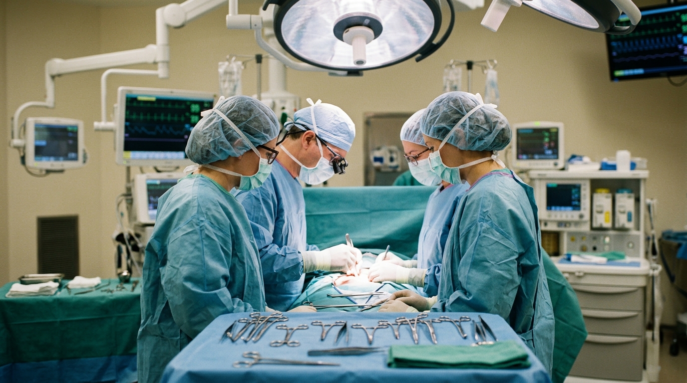
Surgical specialists at Sigmund Freud University Vienna and Wiener Gesundheitsverbund Klinik Floridsdorf have documented the first clinical application of sequential double-layer intraoperative fascial traction to reconstruct an extreme loss-of-domain incisional hernia. The case report, published in the Journal of Abdominal Wall Surgery, describes a thirty-five-year-old male patient who presented with a massive recurrent midline incisional hernia measuring twenty-one centimeters in transverse width following a catastrophic sequence of polytrauma, emergency bowel surgery, open abdomen management, and failed mesh repair. Incisional hernias involving loss of domain represent one of the most formidable dilemmas in reconstructive abdominal surgery because retracted lateral musculature and herniation of a substantial portion of the visceral organs into the hernia sac prevent standard midline closure. Forcibly closing such extensive fascial defects without specialized preparatory adjuncts risks fatal spikes in intra-abdominal pressure and the development of abdominal compartment syndrome. To overcome these anatomical obstacles, the surgical team introduced a staged intraoperative traction protocol utilizing the Fasciotens Abdomen device applied first to the posterior rectus sheath and subsequently to the anterior sheath, evaluating whether mechanical fascial stretching could facilitate layered closure.
The patient’s complex surgical history originated in March 2011 after sustaining severe polytrauma from falling in front of a subway train. His initial traumatic injuries included a pelvic ring fracture, multiple extremity fractures, post-traumatic lower-extremity plexus paresis, serial rib fractures accompanied by right-sided hemothorax, bladder rupture, splenic rupture managed non-operatively, and a ruptured right kidney that mandated an emergency nephrectomy. Severe concurrent transverse colon trauma required a right hemicolectomy with an end colostomy and ileostomy, followed by abdominal closure using a skin flap. Ten years later, in June 2021, the patient suffered an acute abdomen precipitated by an incarcerated incisional hernia with small bowel gangrene. The resulting emergency intervention included para-ileostomy revision, ileo-transversostomy, and open abdomen treatment with negative pressure vacuum-assisted closure. An intraperitoneal onlay mesh repair using a biological graft failed due to subsequent infection requiring explantation, ultimately compelling wound closure by split-skin grafting directly over exposed viscera and leaving a wide epigastric expanse devoid of native peritoneal coverage.
When evaluated for definitive reconstructive surgery, the patient presented with a recurrent midline incisional hernia categorized under the European Hernia Society classification as M1 through M4, W3. Repeated physical examinations revealed superficial wound breakdown along the junction between native skin and the split-skin-grafted sac, managed conservatively with hypochlorous acid wound therapy until stable healing was established. Detailed preoperative planning required precise volumetric assessment through computed tomography to quantify domain loss. Applying the validated ellipsoid geometric formula to axial and sagittal scans demonstrated a hernia sac volume of 6,644 milliliters compared to a residual abdominal cavity volume of 10,053 milliliters. These measurements established a Tanaka index of 0.661, more than 2.6 times the standard clinical diagnostic threshold of 0.25 for loss of domain, alongside a Sabbagh index of 39.8 percent against a twenty percent threshold.
To mitigate these severe physiological and anatomical challenges, the clinical team executed an aggressive multimodal prehabilitation regimen before attempting reconstruction. Subcutaneous tissue expansion was initiated to generate sufficient viable cutaneous coverage to replace the non-preservable skin graft. However, three of the expander pockets contracted intractable infections with Staphylococcus epidermidis resistant to targeted systemic cephalosporin therapy, necessitating their premature surgical removal; only the 800-milliliter right precostal expander remained viable until the definitive operation. Concurrently, chemical component relaxation was induced eight weeks preoperatively by administering ultrasound-guided botulinum toxin A injections totaling three hundred units divided evenly across both lateral abdominal walls, penetrating the external oblique, internal oblique, and transversus abdominis muscles. This prolonged interval extended beyond conventional two-to-four-week windows to coordinate tissue expansion management while maintaining myorelaxation.
Definitive surgical reconstruction was undertaken in March 2025 with the patient under general anesthesia and continuous intra-abdominal pressure monitoring maintained via an indwelling urinary catheter. The procedure commenced with the excision of the remaining right precostal tissue expander and complete removal of the prior split-skin graft, followed by meticulous enterolysis of extensive small bowel adhesions. Retromuscular dissection began cranial to the defect using the Rives-Stoppa technique, integrated with pre-transversalis and pre-diaphragmatic dissection according to the Madrid modification to ensure expansive cephalad mobilization. Bilateral transversus abdominis release was then executed in a cranial-to-caudal direction to release the contracted lateral abdominal wall complexes. Because prolonged open abdomen therapy and previous skin grafting had completely eliminated the peritoneal layer in the upper abdomen, standard midline approximation of the posterior myofascial envelope remained technically impossible despite maximal bilateral transversus abdominis advancement, prompting the team to employ their novel sequential intraoperative fascial traction technique.
The first stage of intraoperative fascial traction targeted the deep myofascial plane using the Fasciotens Abdomen system, which applies controlled mechanical tension to induce viscoelastic stress relaxation and tissue creep. Traction sutures engaged the margins of the posterior rectus sheath extending from the caudal aspect of the incision to eight centimeters inferior to the xiphoid process, maintaining the cranial posterior sheath intact in accordance with Madrid technique guidelines. A sustained diagonal-anterior traction force reaching up to eighteen kilograms was applied continuously for thirty minutes. This targeted mechanical loading lengthened the posterior tissues sufficiently to permit primary approximation and suture closure of the posterior layer from the symphysis pubis up to the level of the umbilicus. Nevertheless, posterior traction could not overcome the complete absence of peritoneal tissue in the epigastrium, leaving a residual defect in the uppermost wound segment where anatomical tissue simply did not exist to bridge the gap.
To establish a durable visceral separation barrier across this remaining cranial gap, surgeons transitioned to the second operative phase by implanting an OviTex S2 permanent reinforced tissue matrix measuring thirty by twenty-five centimeters. The biocomposite graft was tailored to achieve a wide peripheral overlap of at least five centimeters beyond the defect boundaries, secured circumferentially with running continuous sutures to the margins of the mobilised posterior rectus sheath caudally and anchoring directly to the diaphragmatic crura cranially. This reinforced biologic scaffold isolated the underlying peritoneal contents from the overlying retrorectus space, preventing intestinal contact and providing a stable foundation across the epigastric defect.
Following stabilization of the posterior visceral barrier, the surgical team initiated the third phase by repositioning the Fasciotens device to address the outer abdominal wall defect. The anterior rectus sheath presented a persistent midline separation of ten centimeters after posterior closure. Traction sutures were placed along the bilateral anterior fascial edges, and an eighteen-kilogram traction load was applied for an additional thirty minutes. The resulting progressive mechanical creep successfully elongated the anterior rectus sheaths, permitting complete tension-free approximation of the anterior margins across the midline. A large macroporous polyvinylidene fluoride monofilament mesh, DynaMesh-CICAT measuring thirty by thirty-five centimeters, was then positioned within the expanded retromuscular space overlying both the closed caudal posterior sheath and the cranial OviTex visceral bridging graft.
The patient demonstrated an uneventful early recovery, remaining intubated for twenty-four hours before successful extubation and spending two days in the intermediate care unit. Serial bladder pressure measurements confirmed that intra-abdominal pressure remained safely below twelve millimeters of mercury throughout the initial forty-eight postoperative hours, showing no evidence of intra-abdominal hypertension or abdominal compartment syndrome. Closed-incision negative pressure therapy was removed on the fourth postoperative day with primary wound healing noted, allowing hospital discharge on postoperative day ten without systemic complications. At six months postoperatively, computed tomography revealed a seroma that was treated with a single percutaneous aspiration of four hundred milliliters of sterile fluid, with culture showing no bacterial growth. Follow-up computed tomography performed at twelve months postoperatively verified durable abdominal wall restoration without fascial breakdown, mesh displacement, or recurrent fluid accumulation.
From the patient’s subjective perspective, the anatomical restoration produced profound functional improvements in daily life. Before the operation, the individual reported having lost hope for meaningful clinical improvement while living with severe structural instability. Following reconstruction, he reported complete alleviation of chronic lower back pain, significantly enhanced physical mobility, and the capacity to sit comfortably for prolonged periods despite his long-standing post-traumatic extremity paresis. These substantial functional recoveries enabled him to pursue vocational retraining with anticipated workforce reintegration. However, the authors noted several key limitations inherent in their single-case observation. Because the width of the posterior fascial gap prior to traction was not formally measured, the precise quantitative advancement gained through posterior loading cannot be determined. Furthermore, the single-patient design prevents researchers from isolating the relative therapeutic contributions of posterior traction, preoperative botulinum toxin relaxation, and bilateral transversus abdominis release.
The authors concluded that while sequential double-layer intraoperative fascial traction represents a technically feasible addition to the surgical management of massive loss-of-domain incisional hernias, its clinical role requires measured interpretation. The findings demonstrate that posterior traction facilitates substantial fascial mobilization capable of closing inferior portions of the defect, yet it cannot compensate for absolute deficits of peritoneal tissue caused by prior open abdomen therapy, where visceral bridging grafts remain indispensable. The investigators emphasize that prospective clinical series incorporating standardized measurements of fascial gap dimensions, applied traction forces, and physiological airway pressures before and after fascial approximation will be essential to establish the true efficacy of dual-plane intraoperative traction and to identify which patient subsets can achieve complete autologous posterior closure without synthetic or biologic bridging.
Subject of Research: Sequential intraoperative fascial traction for extreme loss-of-domain incisional hernia repair
Article Title: Case Report: Sequential posterior and anterior intraoperative fascial traction in extreme loss-of-domain incisional hernia
Article References: Fortelny, R. H., Froehlich, S., & Kapitanov, T. (2026). Case Report: Sequential posterior and anterior intraoperative fascial traction in extreme loss-of-domain incisional hernia. Journal of Abdominal Wall Surgery, 5, Article 17341. https://doi.org/10.3389/jaws.2026.17341
Image Credits: AI Generated
Keywords: incisional hernia, loss of domain, intraoperative fascial traction, Fasciotens Abdomen, abdominal wall reconstruction, transversus abdominis release, Rives-Stoppa repair, Madrid modification, botulinum toxin A, prehabilitation, tissue expansion, CT volumetry, Tanaka index, OviTex mesh, DynaMesh-CICAT, small-bites technique, intra-abdominal pressure, case report
News Source: Ophelia Keating. (November 13, 2026). Sequential Two-Layer Fascial Traction Closes Severe Abdominal Defect in Clinical Case. Scienmag.