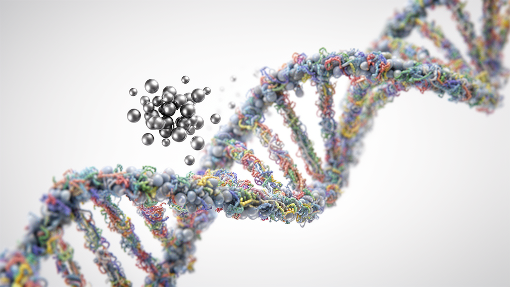
Selenium represents an essential dietary trace micronutrient in human physiology with an exceptionally narrow therapeutic window, where recommended daily intake ranges strictly between 55 and 90 micrograms per day. Clinical and biological investigations demonstrate that falling below this physiological range leads to severe pathologies, including Kashin-Beck disease, an endemic chronic osteoarthropathy marked by the necrosis of growth plates in joint cartilage and bones, alongside increased risks of thyroid dysfunction, impaired fertility, and elevated cancer susceptibility. Conversely, when daily consumption exceeds 400 micrograms, chronic intake rapidly precipitates selenosis, manifested clinically by alopecia, severe nail dystrophy, and, at extreme exposures surpassing 900 micrograms per day, potentially fatal systemic toxicity. To resolve this narrow margin between vital cellular function and lethal poisoning, a comprehensive scientific review published in Health Nanotechnology details how elemental selenium nanoparticles, designated as SeNPs, provide an effective drug delivery and nutrient platform that significantly mitigates systemic toxicity while actively orchestrating complex epigenetic mechanisms across mammalian cells and agricultural plant models.
The physical and biological characteristics of elemental selenium nanoparticles differ fundamentally from dissolved inorganic selenium species, such as selenate and sodium selenite, which carry higher inherent cellular toxicity. In preclinical animal trials, formulating elemental selenium into nanoparticulate suspensions decreases systemic toxicity by up to fourfold compared to conventional aqueous selenite solutions, with hepatic toxicity showing particularly pronounced reductions. Chemical engineering strategies have further enhanced nanoparticle performance by conjugating outer surface coatings with natural polymers and proteins to control physical dimensions and prevent micro-particle aggregation. When synthesized in the presence of bovine serum albumin, the protein binds selenium via cysteine residues to form stable conjugates that exhibit a sevenfold reduction in murine toxicity relative to free selenite, shifting toxic thresholds from 14 milligrams to 113 milligrams of selenium per kilogram of body weight.
Beyond general delivery advantages, functionalized surface modifications enable targeted therapeutic intervention against specific malignant cell lines. Because human tumor types across the liver, kidneys, lungs, and skin frequently overexpress folate receptors, scientists have engineered folic acid-conjugated selenium nanoparticles to achieve selective receptor-mediated uptake. In experimental 4T1 breast cancer cell assays, functionalization with folic acid increased targeted cell mortality by 68 percent relative to unmodified bare nanoparticles, while also triggering apoptotic pathways in HepG2 hepatocellular carcinoma, HK-2 kidney, and MG-63 osteosarcoma cells without generating widespread off-target damage. In vivo murine xenograft models corroborate these in vitro mechanisms, demonstrating that targeted selenium nanoparticle delivery markedly suppresses active tumor growth rates while avoiding the organ damage traditionally linked to high-dose aqueous selenium administration.
At the molecular level, the synthesis and administration of selenium nanoparticles drive extensive remodeling of the epigenetic landscape, transcending classical non-correlated changes in DNA methyltransferase expression. The review identifies that both selenium nanoparticles and aqueous selenite stimulate the transcription of key histone methyltransferase genes, specifically EZH2, SETD7, and SUV39H2, which catalyze the methylation of histone H3 at lysine residues H3K27me3, H3K4me3, and H3K9me3, respectively. Simultaneously, exposure to nanoparticulate selenium represses histone demethylases, including KDM6B, KDM6A, and KDM1B, generating a widespread global increase in H3K4me3, H3K27me3, and H3K9me2 marks alongside reductions in H3K9me3. These transcriptional and enzymatic alterations do not depend on the upregulation of methionine adenosyltransferase 1, whose expression remains unaffected by nanoparticle treatment. Instead, selenium actively participates in one-carbon metabolism, where chemical interactions divert homocysteine into the transsulfuration pathway, producing elevated levels of intermediate selenoglutathione and selenide, thereby clearing the potent catalytic methyltransferase inhibitor S-adenosylhomocysteine and optimizing cellular methylation capacity.
Selenium exposure further modulates post-translational histone acetylation and the expression profiles of non-coding regulatory RNAs that coordinate cellular differentiation and stress responses. Metabolites derived from selenium processing, including seleno-alpha-keto acids formed from Se-methylseleno-L-cysteine, directly inhibit histone deacetylase enzymes due to their structural resemblance to the classical inhibitor butyrate. Building on this inhibitory dynamic, synthetic chemistry has combined a selenium dimer into the therapeutic backbone of vorinostat to create SelSA-1, an agent that binds the catalytic core of multiple deacetylase enzymes with enhanced potency.
The regulation of the human selenoproteome, which comprises twenty-five distinct selenocysteine-containing proteins, constitutes an indispensable axis linking selenium bioavailability to cellular survival, apoptosis, and non-apoptotic cell death. Selenocysteine is inserted cotranslationally into polypeptide chains at designated UGA codons directed by cis-acting insertion sequence elements and specialized transfer RNAs, positioning the reactive chalcogen directly within active catalytic domains. Key oxidoreductases such as glutathione peroxidases and thioredoxin reductases rely on these centers to quench reactive oxygen species and preserve intracellular redox equilibrium. The review underscores that physiological concentrations of selenium nanoparticles support cell cycle progression and inhibit apoptotic pathways, whereas supranutritional doses induce cell cycle arrest and trigger apoptosis in malignant lines. In metastatic lung adenocarcinoma models harboring sensitizing EGFR or KRAS mutations, aqueous selenite acts as a ferroptotic inducer by promoting lipid peroxidation and synergizing with targeted inhibitors like osimertinib and adagrasib, whereas in intracerebral hemorrhage models, selenium nanoparticles prevent ferroptotic neuronal destruction by elevating glutathione peroxidase 4 expression.
Clinical trials and human epidemiological evaluations emphasize that systemic selenium status exerts nuanced, double-edged effects that challenge simplified assumptions regarding broad nutritional supplementation. Observational studies demonstrate clear correlations between low circulating plasma selenium or suppressed selenoprotein P concentrations and elevated risks for hepatocellular carcinoma in Western Europe, as well as higher incidences of thyroid, ovarian, and breast malignancies. Nevertheless, large-scale randomized human trials demonstrate significant caveats: the landmark Selenium and Vitamin E Cancer Prevention Trial demonstrated that dietary supplementation with L-selenomethionine failed to prevent prostate cancer in men with low baseline selenium, while significantly increasing high-grade prostate cancer risk in individuals possessing high endogenous selenium reserves.
Epigenome-wide association studies across human populations have validated that circulating selenium concentrations and dietary intake directly correlate with distinct DNA methylation patterns in functional metabolic pathways. In the Obekit clinical study evaluating 337 participants undergoing dietary intervention, specific nutritional selenium intake correlated with altered CpG methylation at cg16682935 within the PAPSS2 gene, an integral component of the physiological sulfate activation cascade. Furthermore, clinical investigations involving obese women linked depressed serum selenium levels to site-specific hypermethylation of CPT1B, a gene governing mitochondrial fatty acid beta-oxidation. Research examining American Indian cohorts identified five distinct differentially methylated positions linked to dietary selenium, prominently featuring hypermethylation at cg00163554 within the DIP2C locus, an enzyme whose genetic disruption induces broad genomic hypomethylation. In neonatal cohorts, higher measured placental selenium concentrations correlated with elevated hypertonicity and site-specific hypermethylation mapping directly to GFI1, revealing that prenatal maternal trace element exposure actively influences fetal neurodevelopmental trajectories.
In agricultural systems and environmental plant biology, selenium is not classified as an essential nutrient, yet soil availability fundamentally dictates plant physiology, crop stress tolerance, and human dietary intake. Plants absorb inorganic selenium primarily as selenate through root sulfate transporters under alkaline, oxidizing conditions, whereas selenite predominates in neutral to acidic soils where it strongly adsorbs to iron oxides, limiting bioavailability. Agricultural crops accumulate selenium, converting it into organic selenocysteine and selenomethionine, which provide the primary dietary source for grazing livestock and human populations. While moderate selenium exposure mitigates abiotic stresses like drought and excessive soil salinity, elevated tissue concentrations generate toxic reactive oxygen species and impair nutrient absorption. In Capsicum annuum, excessive in vitro applications of 10 to 45 nanometer elemental selenium nanoparticles above 10 milligrams per liter triggered aberrant DNA methylation changes that disrupted apical meristem development, highlighting the critical necessity for precise dose control during agronomic biofortification initiatives.
Addressing modern agricultural challenges is becoming increasingly urgent because global climate change and declines in anthropogenic coal combustion emissions are projected to deplete soil selenium concentrations across extensive cropland regions in Europe, North America, Southern Africa, and India. With an estimated 500 million to one billion people currently suffering from suboptimal selenium intake or frank deficiency, advanced biofortification methodologies, ranging from foliar applications to genetic interventions, are essential to maintain food security. To monitor these dynamic biochemical transformations, researchers deploy advanced analytical technologies, including High Energy Resolution Fluorescence Detected X-ray Absorption Spectroscopy and Nanoscale Secondary Ion Mass Spectrometry. Operating at cryogenic temperatures of 10 Kelvin to eliminate beam radiation damage, fluorescence-detected spectroscopy achieves resolution down to 400 micrograms per kilogram in biological tissue, enabling researchers to track nanoparticle transformations, quantify organic selenocompounds, and design safe, targeted selenium nanotechnologies for human therapeutics and global crop enrichment.
Subject of Research: Epigenetic regulation and reduced toxicity profiles of selenium nanoparticles in human and plant systems
Article Title: Selenium nanoparticles and the epigenetic landscape: emerging mechanisms in human and plant health
Article References: Conlan, R. S., Toubhans, B., & Charlet, L. (2026). Selenium nanoparticles and the epigenetic landscape: emerging mechanisms in human and plant health. Health Nanotechnology, 2(1), Article 11. https://doi.org/10.1186/s44301-026-00034-8
Image Credits: AI Generated
DOI: 10.1186/s44301-026-00034-8
Keywords: selenium nanoparticles, epigenetics, DNA methylation, histone methyltransferases, selenoproteins, glutathione peroxidase 4, ferroptosis, chitosan coating, bovine serum albumin, folic acid targeting, transsulfuration pathway, Kashin-Beck disease, biofortification, HERFD-XAS, NanoSIMS, thioredoxin reductase, environmental selenium
News Source: Juliet Wilcox. (November 18, 2026). Selenium Nanoparticles Modulate Epigenetic Pathways While Reducing Toxicity in Biological Systems. Scienmag.