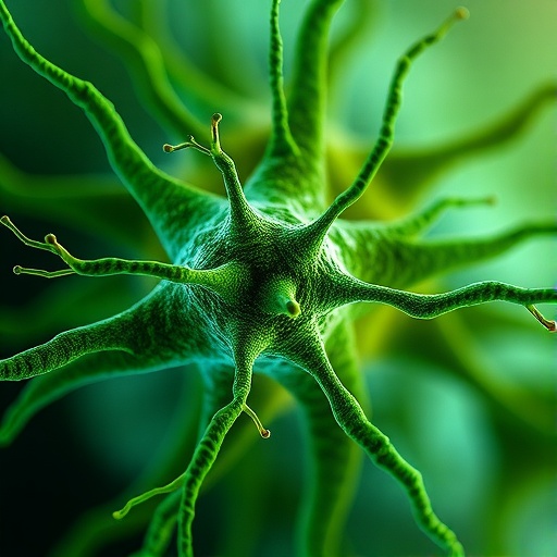
A groundbreaking study by researchers at the German Center for Neurodegenerative Diseases (DZNE) uncovers the internal cellular mechanism that determines why a neuron typically extends only one axon—a critical feature for proper brain function. Challenging the dominant theory that external environmental cues dictate axon formation, this research reveals that the axon’s emergence is driven primarily by intrinsic activity within the neuron itself.
Neurons initially exhibit symmetrical small projections called neurites during early development. Traditionally, it was believed that external growth factors selectively stimulate one neurite to transform into the axon. However, this new study, published in Nature, demonstrates that the process is governed by cyclic remodeling of the cytoskeleton originating within the neuron’s soma, or cell body. This intrinsic process disrupts the symmetry by enabling one neurite to stabilize and elongate, eventually becoming the singular axon.
Researchers observed that young neurons undergo rhythmic behavior characterized by repetitive extension and partial retraction of their neurites—akin to “two steps forward and one step back.” This oscillatory pattern unfolds over about 48 hours, culminating in the persistent growth of a single neurite. The others later mature into dendrites, specialized to receive incoming signals.
The core driver of this rhythmic cellular “shape-shifting” is a protein complex known as Arp2/3. This molecular complex functions like a zipper, locally loosening the cytoskeletal “corset” that otherwise maintains the neuron’s rigidity. By transiently relaxing the cytoskeletal meshwork in a wave-like propagation along one neurite at a time, Arp2/3 enables episodic growth spurts. These waves of remodeling continue until internal forces within the cytoskeleton reestablish tension, causing the projection to shrink again.
Interestingly, this process shows a stochastic element: the wave can reoccur on the same or a different neurite. Over time, one neurite gains stability through the internal growth of stiff structural proteins, allowing it to resist retraction and extend independently of the Arp2/3-driven oscillations. Once this neurite commits to axonal growth, the rhythmic waves subside, cementing neuronal polarity.
While external factors cannot be fully excluded, the study strongly implicates neuron-intrinsic cytoskeletal oscillators as the primary architects of axon specification. How this oscillatory remodeling is genetically programmed, why it is rhythmic, and how one neurite ultimately dominates remain open questions. The findings invite further research into the genetic and molecular pathways that coordinate these fundamental cytoskeletal dynamics.
Overall, this discovery reshapes our understanding of neuronal polarization, revealing that the precise wiring of the brain is sculpted not just by signals from outside the cell, but through carefully orchestrated internal mechanical rhythms. These insights hold promise for advancing therapeutic approaches to neurodegenerative diseases by targeting intrinsic cellular mechanisms underlying neuron development and function.
Subject of Research: Cells
Article Title: An intrinsic cytoskeletal oscillator establishes neuronal polarity
News Publication Date: 8-Jul-2026
Web References: http://dx.doi.org/10.1038/s41586-026-10755-6
References: Lin, T.-C., et al. (2026). An intrinsic cytoskeletal oscillator establishes neuronal polarity. Nature.
Keywords: Neuroscience, Cell biology, Molecular biology
Tags: cytoskeleton remodeling in neuronsinternal cellular processes in neurogenesisintrinsic neuron activity in nerve cell developmentneurite outgrowth regulationneurodevelopmental mechanisms governing axon emergenceneuron polarity establishment during developmentneuron soma cytoskeleton dynamicsneuron symmetry breaking mechanismneuronal axon formationrhythmical neurite extension and retractionrole of protein complexes in nerve cell growthsingle axon determination in neurons