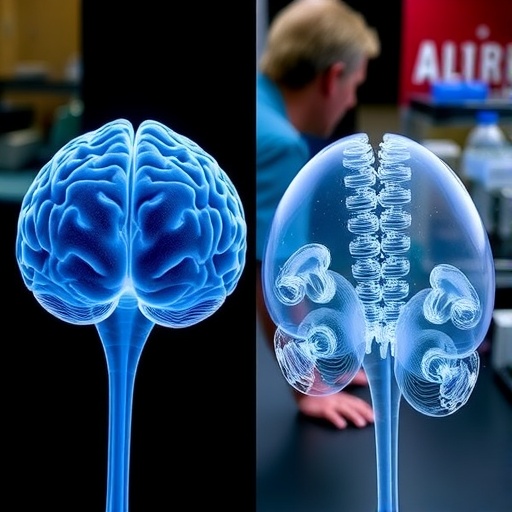
For nearly 15 years, scientists have been building organoids—three-dimensional structures grown from stem cells that mimic selected features of organs in miniature. Researchers at the Institute of Science and Technology Austria (ISTA) have now developed a robust mouse cortical organoid model that reproduces many of the cellular and molecular characteristics of the developing cerebral cortex. The study, published in Nature, offers a powerful new way to examine how the brain forms, how it reaches the correct size, and why disruptions in development can produce conditions such as microcephaly or macrocephaly. At the same time, the work reveals a crucial limitation: although the organoids generate many of the same cell types as a living mouse brain, the sequence and timing of development are not fully synchronized.
Organoids are produced from stem cells, which can both renew themselves and generate specialized descendants, including neurons and glial cells. Under carefully controlled conditions, these cells can organize themselves into structures that resemble aspects of developing tissues. Brain organoids are particularly valuable because they allow researchers to study early neural development in a three-dimensional environment outside the body. However, most established systems have been derived from human stem cells, making them useful for investigating human biology but often difficult to manipulate genetically. Mice, by contrast, offer a much larger collection of genetic tools for tracing individual cells, altering specific genes, and linking cellular behavior to developmental outcomes. The ISTA team set out to combine the organizational advantages of organoids with the experimental precision of the mouse model.
The project was led by the Hippenmeyer group, with Melissa Stouffer, Osvaldo Miranda Romero, Florian Pauler, and Fabrizia Pipicelli as co-lead authors, alongside Carmen Streicher and Giselle Cheung. Developing the system required more than simply placing mouse stem cells in a culture dish. The researchers first established a stable mouse stem cell line and then optimized a reproducible protocol capable of generating cortical organoids across different experimental batches and cell lines. Stouffer trained in advanced three-dimensional brain organoid methods at Stanford University’s Brain Organogenesis Workshop, an experience that helped the team address the technical challenges of maintaining stem cells and producing consistently structured organoids. This groundwork was essential because variability between organoids can obscure genuine biological patterns and make developmental comparisons difficult.
The resulting structures contained cortical rosettes, organized formations that resemble aspects of the neural tissue found in the developing cerebral cortex. These rosettes appeared as dense regions along the outer edge of the organoids and were composed of neural progenitor cells, the descendants of stem cells that generate the brain’s neurons and glial cells. To test how faithfully the organoids modeled development, the researchers compared them directly with developing mouse brains rather than relying on comparisons between different species. They examined corresponding developmental stages using single-cell sequencing, a method that measures gene activity in individual cells. This approach allowed the team to determine which cell populations were present, how abundant they were, which molecular programs were active, and when particular cell types emerged or declined.
At a broad level, the organoids closely resembled the developing mouse cortex. The researchers detected similar populations of neural progenitors, neurons, and glial cells in both the organoids and living tissue. They also found that many of the molecular programs guiding development were shared. This similarity means that mouse cortical organoids can provide meaningful information about fundamental mechanisms of brain formation, including the behavior of radial glia. Radial glial cells are neural stem and progenitor cells that both produce new brain cells and provide structural support during cortical development. Their descendants eventually form the layered architecture of the cerebral cortex, a region essential for sensory processing, movement, learning, and cognition.
The most striking difference emerged when the team examined developmental timing. In the living mouse brain, cortical development follows a largely ordered sequence. Neural stem cells initially expand through symmetric divisions, in which one cell produces two similar progenitor cells. They then shift toward asymmetric divisions, generating one progenitor and one differentiating daughter cell, a transition that supports the production of neurons. After the major wave of neuron formation has ended, the production of glial cells becomes prominent. These phases are arranged in a temporal progression, allowing the developing brain to coordinate cell production with tissue growth and organization.
In the cortical organoids, the same broad cellular identities appeared, but the schedule was less tightly controlled. Neural development became temporally uncoupled: populations and programs that normally arise in sequence could overlap or appear at different relative times. The organoids preserved the overall “hour” of development but not its precise “minutes,” as the researchers describe it. The emergence of glial cells was a notable exception. In both the organoids and the mouse brain, glial development followed neuron formation, suggesting that some parts of the developmental timetable are intrinsically robust while others depend more heavily on signals from the surrounding organism. This distinction could be critical when interpreting organoid experiments involving disease-associated genes or developmental disorders.
To investigate lineage progression in greater detail, the researchers used MADM, or Mosaic Analysis with Double Markers. This genetic technique labels cells and their descendants with different fluorescent markers after specific chromosome-segregation events, enabling scientists to follow the behavior of individual progenitor cells and their lineages. The method has previously allowed the Hippenmeyer group to map the development of the mouse cortex at the level of single progenitor cells. Applying the same conceptual framework to organoids made it possible to compare how stem cells divide and produce descendants in a living brain versus an isolated three-dimensional culture. The findings indicate that self-organization alone can reproduce much of the cortical program, but cannot fully impose the orderly progression observed in vivo.
The likely explanation is the absence of the stem-cell niche. In the living brain, neural progenitors do not develop in isolation. They interact with neighboring cells, blood vessels, signaling molecules, growth factors, extracellular structures, and mechanical forces. These external inputs can influence when stem cells divide, when they switch from producing neurons to producing glia, and how newly generated cells are positioned. Organoids contain some internal communication and can organize themselves without direct instruction, but they lack many of the systemic signals supplied by an intact organism. The study therefore identifies not only what cortical organoids can reproduce, but also which developmental processes remain dependent on environmental context.
The new model gives researchers a controlled platform for testing how genetic mutations, biochemical signals, or physical conditions affect cortical development. It may help clarify why changes in the regulation of neural stem cells lead to unusually small or large brains and could reveal which developmental stages are most vulnerable to disruption. The researchers’ next goal is to recreate selected components of the stem-cell niche within the organoids by adding factors found in the mouse brain. If those interventions restore a more linear developmental sequence, they could improve the precision of organoid models and make them more useful for studying neurological disease. The work establishes a clear benchmark for future organoid research: similarity to a real brain should be measured not only by the cell types present, but also by the timing, lineage relationships, and environmental signals that bring those cells into existence.
Subject of Research: Lab-produced tissue samples
Article Title: Temporal Uncoupling of Radial Glia Lineage Progression in Cortical Organoid
Web References: Nature article; ISTA Hippenmeyer Group
References: Nature; DOI: 10.1038/s41586-026-10916-7
Image Credits: © Stouffer et al. / Nature
Keywords
Cortical organoids, brain organoids, mouse models, cerebral cortex, neural stem cells, radial glia, brain development, neuroscience, single-cell sequencing, MADM, tissue cultures, organ development
Tags: brain organoid limitationsbrain organoidscerebral cortex developmentearly neural development in vitromouse cortical organoidsneural cell type differentiationneurodevelopmental disorder researchorganoid comparison to human brainsorganoid modeling of neurodevelopmental conditionsstem cell-derived brain modelsstudying brain size abnormalitiesthree-dimensional brain tissue models