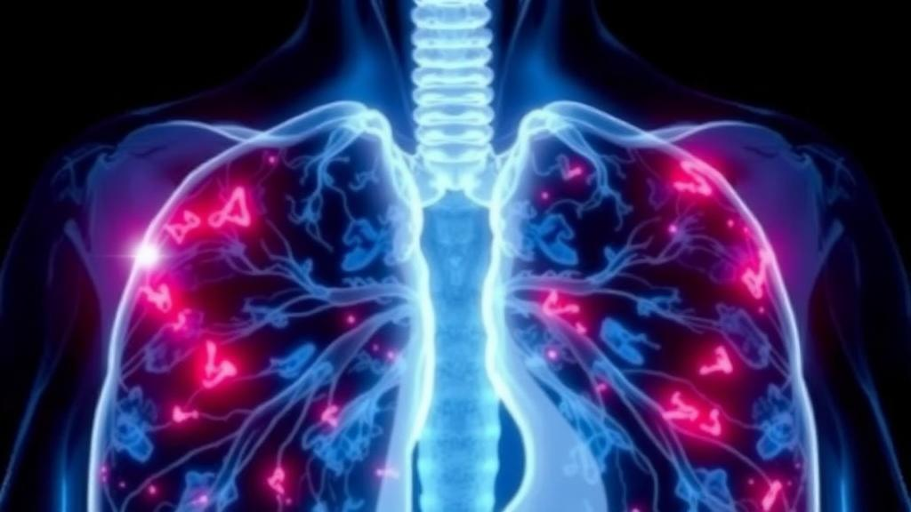
Idiopathic pulmonary fibrosis has long been one of medicine’s most frustrating adversaries, a disease in which the lungs stiffen and scar for reasons that remain stubbornly unclear, and in which two patients who look identical on paper can follow wildly divergent paths. One may remain stable for years while another deteriorates rapidly toward respiratory failure. Now a team of researchers at the Medical University of Gdańsk in Poland reports that a standard nuclear medicine scan may be able to see that difference coming. In an exploratory retrospective study published in BMC Medical Imaging, the investigators found that the intensity of glucose metabolism inside scarred lung tissue, measured with fluorodeoxyglucose positron emission tomography combined with computed tomography, rises in step with established clinical measures of disease severity, and may even foreshadow how quickly the disease will progress.
The clinical benchmark for staging idiopathic pulmonary fibrosis is the GAP score, an acronym drawn from the variables it incorporates: gender, age, and physiology, the latter captured through lung function tests such as forced vital capacity and the transfer factor for carbon monoxide. The GAP score is a powerful and convenient tool, but it is fundamentally static. It describes the damage that has already accumulated; it says nothing about the biological activity of the disease at the level of the tissue itself. That distinction matters because fibrosis is not merely a passive scar. In active disease, fibroblasts proliferate, inflammatory cells infiltrate, and remodeling processes consume energy, and all of that cellular work demands fuel, principally glucose.
This metabolic hunger is precisely what fluorodeoxyglucose exploits. The radiotracer is a glucose analog labeled with fluorine-18, and once injected it is taken up by metabolically active cells, where it becomes trapped after phosphorylation and emits positrons that the PET scanner detects. In oncology, this principle underpins the familiar maximum standardised uptake value, or SUVmax, a number that quantifies the hottest spot of tracer accumulation within a lesion. The Gdańsk team asked a deceptively simple question: could the same measurement, applied not to tumors but to fibrotic lung parenchyma, serve as a window into the aggressiveness of pulmonary fibrosis?
To find out, the researchers turned to a cohort of thirty patients with idiopathic pulmonary fibrosis who had undergone fluorodeoxyglucose PET/CT for suspected comorbid disease, most often to rule out malignancy. This is a common clinical scenario, since patients with pulmonary fibrosis carry an elevated risk of lung cancer, and the retrospective design allowed the team to mine scans that had already been acquired for other reasons. The researchers measured raw SUVmax values within the fibrotic regions of the lung and compared those values against each patient’s GAP score calculated at the time of the scan. Because uptake values and severity scores are not normally distributed in a sample of this size, they used Spearman’s rank correlation coefficient, a non-parametric statistic that assesses whether two variables tend to increase together without assuming a linear relationship.
The answer was a qualified yes. The correlation between GAP score and fibrotic-lung SUVmax was positive and statistically significant, with a Spearman coefficient of 0.448 and a p-value of 0.013. In plain terms, patients whose lungs burned through more glucose on the scan tended to be the patients whose disease was already more advanced by conventional clinical criteria. A coefficient of 0.448 represents a moderate association, not a perfect one, and the authors are careful to frame the finding as exploratory. Yet the implication is striking: a molecular imaging measurement, taken from a single scan session, appears to carry information that overlaps with, and potentially extends beyond, the physiologic staging that clinicians currently rely upon.
The more provocative part of the study concerns outcomes. The researchers split their cohort into high and low metabolic activity groups using a median split of the SUVmax values, set at 2.53 for the full cohort of thirty patients and at 2.75 for the nineteen patients who were receiving antifibrotic therapy, the modern standard of care with drugs such as the tyrosine kinase inhibitor nintedanib. They then compared progression-free survival and overall survival between the groups using the Mantel-Cox log-rank test, the standard survival analysis method that compares the hazard of an event over time between two populations. In the antifibrotic-treated subgroup, patients with high uptake in their fibrotic lesions showed a median progression-free survival of twenty-five months, exactly half the fifty months observed in the low-uptake group, corresponding to a hazard ratio of 3.68.
Here the statistical caution becomes essential. The confidence interval for that hazard ratio stretched from 0.74 to 18.43, a range that crosses the value of 1.0, meaning the result cannot be distinguished from chance, and the log-rank p-value of 0.089 fell short of the conventional threshold of 0.05. The authors are transparent about this: the survival signal is a hypothesis-generating observation, not a proven predictor. But the pattern is biologically coherent and clinically tantalizing. A hazard ratio approaching four, even with wide uncertainty, suggests that with a larger sample the association could harden into something actionable. If metabolic activity in fibrotic lung truly doubles the pace of progression despite antifibrotic drugs, that information could reshape decisions about monitoring frequency, transplant referral timing, and enrollment into clinical trials of new therapies.
The study’s limitations are the natural consequences of its design. Thirty patients is a small sample, and the scans were acquired for indications unrelated to fibrosis staging, which introduces selection bias even as it offers a glimpse of real-world imaging. The retrospective framework, approved by the Bioethics Committee of the Medical University of Gdańsk with the consent requirement waived, means the researchers could not standardise when or why each scan was performed. SUV measurements are also sensitive to technical factors such as patient blood glucose levels, uptake time, and scanner calibration, all of which complicate comparisons across individuals. The authors themselves conclude that larger prospective studies are required before molecular imaging can be declared an additive prognostic tool in idiopathic pulmonary fibrosis.
Even so, the conceptual advance is worth savoring. For decades, the assessment of fibrotic lung disease has rested on structural and functional endpoints: how much the lung can hold, how well oxygen crosses the membrane, how the tissue looks on high-resolution CT. These measures capture the aftermath of disease. Metabolic imaging, by contrast, offers something closer to a live readout of the pathological process itself, the cellular furnace that drives scarring forward. Previous research has linked fluorodeoxyglucose uptake in the lung to inflammatory and proliferative activity in interstitial disease, and the Gdańsk findings now tie that signal to a validated severity index and to survival trajectories in a treated population.
The road from exploratory correlation to clinical utility is long, and most imaging biomarkers that show promise in small retrospective series never reach the clinic. But the logic of this study points toward a future in which a patient’s PET scan does more than exclude cancer. It could quantify how ferociously their fibrosis is burning, stratify them into risk groups that GAP scores alone cannot resolve, and identify the patients most likely to benefit from intensified therapy or novel antifibrotic agents. For a disease that kills most of those it afflicts within a few years of diagnosis, any tool that reveals the fire beneath the scar, before the lung has irreversibly stiffened, would represent a genuine leap forward. This study is an early, cautious step in that direction, and its authors have mapped the path that the next, larger studies must walk.
Subject of Research: Association between fibrotic-lung FDG-PET uptake and disease severity and outcomes in idiopathic pulmonary fibrosis
Article Title: Association of fibrotic-lung 18F-FDG uptake with GAP severity and clinical outcomes in idiopathic pulmonary fibrosis: an exploratory retrospective study
Article References: Ward, C., Oppmann-Ward, A., Wilczynski, S., Korneliussen, J., Żegleń, S., Rogoza, K., & Cytawa, W. (2026). Association of fibrotic-lung 18F-FDG uptake with GAP severity and clinical outcomes in idiopathic pulmonary fibrosis: an exploratory retrospective study. BMC Medical Imaging. https://doi.org/10.1186/s12880-026-02856-5
Image Credits: AI Generated
DOI: 10.1186/s12880-026-02856-5
Keywords: idiopathic pulmonary fibrosis, FDG-PET/CT, SUVmax, GAP score, pulmonary fibrosis, nuclear medicine, interstitial lung disease, progression-free survival, antifibrotic therapy, molecular imaging, lung fibrosis, prognosis
News Source: Ophelia Keating. (October 6, 2026). Scarring Lungs Light Up on PET Scans, Hinting at How Deadly Pulmonary Fibrosis Will Become. Scienmag.