Charcoal is usually treated as the end of a tree’s story, a fuel lump stripped of everything that once made it wood. A study published in Applied Microscopy on 3 March 2026 argues otherwise. Chul Jong Yoon and Ki Woo Kim fractured pieces of commercial white charcoal with nothing more elaborate than a razor blade and a hammer, then examined the freshly exposed surfaces with a scanning electron microscope. What they found inside was startlingly intact: vessels, fibers, parenchyma cells and pits of the secondary xylem had survived the kiln, and threaded among them were fungal hyphae that had proliferated, branched and even sporulated on the vessel walls. The structures closely resembled those of a fungus known to cause oak wilt disease in South Korea, suggesting the tree behind the charcoal may have been diseased long before it ever reached the fire.
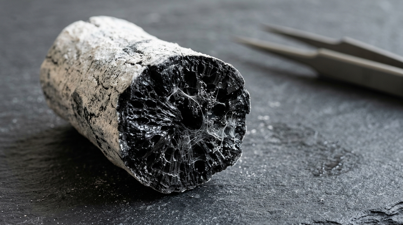
The charcoal in question was not laboratory-made. Three lumps, each roughly 15 centimeters in diameter, were obtained from a wood artisan in Korea. They were presumed to have been made from oak species in 2005 and had been purchased for use in decorative wood crafts. The lumps appeared white to pale gray overall and, when two were knocked together, produced a metallic sound. That whiteness is not cosmetic. White charcoal belongs to a category of charcoal produced at temperatures above 1,000 degrees Celsius, distinct from black charcoal made below 400 degrees and activated charcoal made between 400 and 800 degrees. More specifically, white charcoal is produced by pyrolyzing wood at roughly 200 to 400 degrees Celsius for several days, with the temperature then raised above 1,000 degrees toward the end of the process. The name itself comes from the practice of quenching the hot wood with ash, which imparts a pale gray hue to the surface.
In South Korea, 90 percent of all white charcoal is produced in Gangwon Province from oak trees, primarily the Chinese cork oak, Quercus variabilis, and the material is used for crafts, water treatment and various other daily applications. The wood’s electrical resistivity decreases as carbonization temperature rises, likely because ultrafine graphite-like crystallites form, accompanied by the appearance of sodium compound crystals. That conductivity makes white charcoal a candidate electrode material for fuel cells or batteries. Yet despite several earlier electron-microscopic examinations of white charcoal morphology, the authors noted that little information existed about how to cleave or fracture the material to expose its internal anatomy, and visual evidence of microbial behavior inside the charcoal matrix was scarce. Their stated objective was to assess one such cleaving technique for characterizing microbial behavior within the charcoal via scanning electron microscopy.
The technique was deliberately crude. At ambient temperature, a razor blade was inserted into each air-dried charcoal lump as a makeshift chisel and struck with a hammer, splitting the lump into smaller pieces. The method’s virtue was that it revealed internal surfaces with no need for any polishing or smoothing procedure. Fractured pieces of roughly 1 cubic centimeter were mounted on aluminum stubs with double-sided copper tape, without chemical fixation, ethanol dehydration or critical point drying, and coated with gold using an ion coater. The specimens were then observed in a scanning electron microscope operated at 20 kilovolts. Measurements of vessels and pits were taken, with means and standard deviations calculated in spreadsheet software.
The transverse surfaces that resulted showed heartwood anatomy in remarkable detail. Vessels were concentrated in the earlywood, and nearly all of them were embolized, meaning their lumens were blocked rather than open to water transport. They were predominantly ellipsoidal, with a mean diameter of 252.82 plus or minus 27.09 micrometers across 20 measured vessels. The embolized vessels contained tyloses, balloon-like ingrowths of adjacent parenchyma cells that pass through pits into vessel lumens, and most of these had ruptured during charcoal production, though some retained near-intact membranous forms. Clusters of axial parenchyma cells sat between the larger vessels. On the fracture surface of the transition zone between heartwood and sapwood, embolized and unoccluded vessels coexisted in an approximately 1:1 ratio. Heartwood vessels were blocked by tyloses, while sapwood vessels remained conductive, with open lumens and clearly discernible corrugated secondary cell wall surfaces.
Other xylem components fared equally well under the microscope. Fiber-tracheids appeared with narrower diameters of about 5 micrometers and thicker cell walls of about 2 micrometers than the adjacent axial parenchyma cells, and the inner surfaces of pits were discernible within them. Cell-to-cell junctions between neighboring parenchyma cells were visible in magnified views. On radial fracture surfaces, vessels and rays stood out clearly, and several simple perforation plates could be seen within the vessels. The secondary cell walls of the vessels carried a high density of oval-shaped pits, which averaged 3.14 plus or minus 0.5 micrometers in length across 20 measurements. Both open pits and embolized pits filled with ray cells of varying diameters were observed. Taken together, the presence of ring-porous wood, axial parenchyma cells and xylem fibers suggests the charcoal was produced from a Quercus species, an identification reinforced by the fact that most South Korean white charcoal comes from this genus. The authors caution that the charcoal was not stained for histological analysis, so structures were identified only partially, based on cell morphology and wall thickness in the electron micrographs.
The most striking discovery came from the fungal structures. Hyphae were found adjacent to a membranous tylose within a vessel, although none were observed to pass through the tylose itself. Elsewhere, hyphae proliferated, branched and sporulated on the secondary cell walls of vessels. A magnified view revealed a conidiogenous cell bearing a flat scar, or annellation, and the conidium it produced was oval-shaped and measured approximately 3 micrometers in length. Hyphae were also found within the pits of the secondary cell walls, where the pit apertures appeared large enough to permit the passage and lateral migration of hyphae from one vessel into its neighbors. The authors interpret this as a mechanism for hyphal colonization and spread within the host, a behavior consistent with earlier observations of hyphae entering bordered vessel pits in Norway spruce heartwood after incubation with a white rot fungus.
The morphology of these fungal structures carries a specific identification. Conidiogenous cells with flat scars and obovoid to pyriform conidia measuring approximately 4 to 10 micrometers in length are the described characteristics of Raffaelea quercus-mongolicae, a pathogen known to cause oak wilt disease in South Korea. The conidium observed in the charcoal, at roughly 3 micrometers, sits near but below the lower end of that published range, and the authors describe the match as morphological rather than molecular. Oak wilt disease was first reported in Korea in 2004 and occurs most frequently in Mongolian oak. The number of trees killed annually by the disease peaked at approximately 331,000 in 2011 before declining to about 157,000 in 2019. Given that the wood was harvested in 2005, the authors hypothesized that the trees used for the charcoal had been infected by the pathogenic fungus in the stand, triggering tylosis formation as a defensive response. Because oaks displaying wilt symptoms in the field may still be harvested for construction timber, mushroom cultivation substrate or charcoal production, the observations imply that the oak used here may have been diseased.
Timing matters in this interpretation. The presence of fungal hyphae within the charcoal suggests the fungi had already colonized the wood before carbonization. Post-production colonization by saprophytes, wood-decay fungi or charcoal-degrading fungi can frequently occur on the surface of white charcoal, but invasion after production is considered unlikely here, because the occluded vessels and tyloses act as a physical barrier. The authors therefore reason that fungal colonization occurred prior to carbonization, with distinct morphological details retained through the high-temperature process. The tyloses themselves appear to have served as a physical barrier deterring the spread of hyphae within the vessels, and the presence of ray cells within the pits of embolized vessels likely marks an initial stage of tylosis budding that can lead to complete vessel occlusion. Similar tylosis buds have been identified in Quercus variabilis charcoal and in Eucalyptus globulus chips in earlier work, although the mechanisms by which nascent buds merge and enlarge within vessels remain to be fully elucidated in oak species.
The broader significance lies in what charcoal might remember. Oak species, with their anatomically distinct growth rings and wide ecological tolerance, are commonly used in dendrochronological analyses, and the authors propose that oak charcoal could serve as a proxy for inferring historical pathological events in forests. Xylem embolism has been documented in a range of oak species using fluorescence microscopy, X-ray micro-computed tomography and bright-field microscopy, and vessel occlusion through tylosis formation in response to fungal infection has been observed in infected oaks, findings that corroborate the present results. The authors are careful about the limits of their evidence: the identification relied solely on morphological fungal features, so DNA sequence-based studies are essential to validate the host-parasite interaction and strengthen any pathological inference. They also note that further investigation is needed to track the production history of white charcoal and build a chronological foundation for dendrochronology in Korea. Even so, the study demonstrates that a razor blade, a hammer and a scanning electron microscope can turn a lump of craft charcoal into a readable archive of a tree’s final years, preserving both the wood’s architecture and the traces of the microbe that may have helped fell it.
Subject of Research: Scanning electron microscopy of fractured white charcoal revealing preserved wood anatomy, vessel embolism and fungal structures within the secondary xylem
Article Title: Fractography of white charcoal reveals past fungal infection and embolism in the secondary xylem via SEM
Article References: Yoon, C. J., & Kim, K. W. (2026). Fractography of white charcoal reveals past fungal infection and embolism in the secondary xylem via SEM. Applied Microscopy, 56(1), Article 5. https://doi.org/10.1186/s42649-026-00126-w
Image Credits: AI Generated
DOI: 10.1186/s42649-026-00126-w
Keywords: white charcoal, scanning electron microscopy, secondary xylem, oak, tyloses, xylem embolism, fungal hyphae, Raffaelea quercus-mongolicae, oak wilt, Quercus variabilis, dendrochronology, wood anatomy
News Source: Kristina Jarvis. (November 5, 2026). Scanning Electron Microscope Reveals Fungal Infection Hidden Inside White Charcoal. Scienmag.