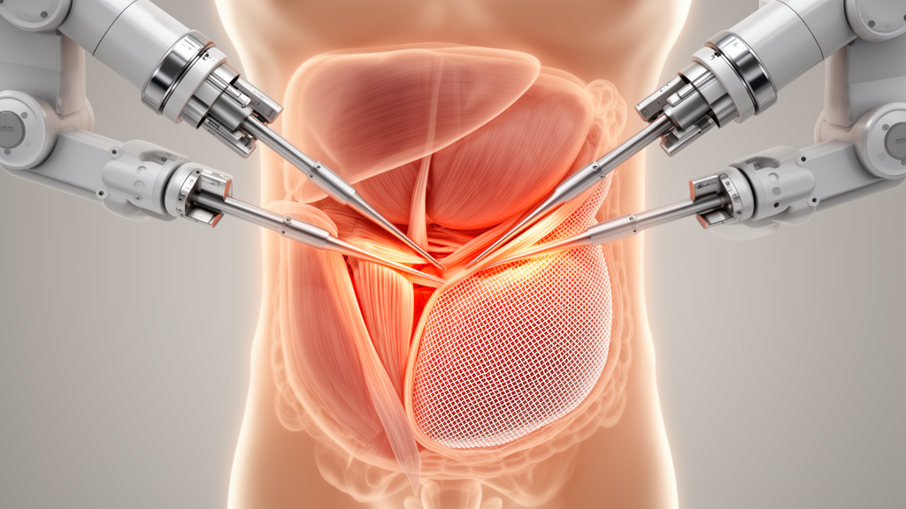
Surgeons in India have reported the first use of intraoperative fascial traction inside a robotic enhanced-view totally extraperitoneal hernia repair, a combination that allowed six patients with very large ventral hernias to leave the operating room with their abdominal midline closed and without the aggressive muscle-cutting procedures that such repairs usually demand. The technique, described in the Journal of Abdominal Wall Surgery, pulled the stretched edges of a hernia defect measuring on average 15.8 centimetres down to 8.9 centimetres during surgery, a mean medialisation of 6.9 centimetres, enough to bring the two halves of the abdominal wall together with minimal tension in every case.
The challenge the team set out to solve is one of the most stubborn in abdominal wall surgery. When a ventral or incisional hernia grows large, particularly in patients with loss of domain, in which a substantial volume of intestine has chronically herniated into the hernia sac and the abdominal cavity has permanently shrunk to accommodate it, simply sewing the fascial edges together generates excessive tension. That tension drives recurrence, wound breakdown, pain, and postoperative complications. Surgeons have traditionally reached for adjuncts such as progressive preoperative pneumoperitoneum, in which air is gradually insufflated into the abdomen over weeks to stretch the cavity, botulinum toxin A injections that paralyse the lateral abdominal wall muscles for months and allow passive lengthening, or component separation, in which the layers of the abdominal wall are surgically divided so the muscles can slide toward the midline.
Each of those strategies carries a price. Pneumoperitoneum demands extended preoperative preparation. Component separation increases surgical complexity and is associated with seroma formation, denervation of the abdominal wall, functional impairment, mesh infection, and long-term hernia recurrence. Botulinum toxin requires a four-week wait and adds cost. Intraoperative fascial traction offers a different philosophy: rather than cutting or chemically relaxing anything, it mechanically elongates the myofascial structures with a controlled, symmetrical stretching force, exploiting the viscoelastic properties of living tissue to lengthen the lateral muscle complex and medialise the rectus muscles while preserving the structural integrity of the abdominal wall.
Until now, that philosophy had been demonstrated almost exclusively in open surgery, where devices can be clamped directly onto the fascia. Only a single case report, by Bloemendaal and colleagues in 2022, had described fascial traction inside a robotic repair, and that was in a transabdominal retromuscular approach using an intra-abdominal technique. The Delhi team, led by Vivek Bindal, Dhananjay Pandey, Deepak Kumar, Shahiq Ahmed, and Aakash Patel at Max Super Specialty Hospital, applied traction within the extraperitoneal eTEP space, a technically distinct plane that never enters the abdominal cavity, using a different port strategy and suture methodology. It is, by their account, the first reported use of fascial traction in a robotic eTEP approach.
The technical execution is intricate. All six operations were performed on the Da Vinci Xi robotic system between June 2024 and March 2025, using the Fasciotens Hernia System to generate the traction. Patients were positioned supine with the left arm tucked to optimise exposure, and the operating table was adjusted so the robotic arms could reach without colliding with the traction frame. Access to the retromuscular space came through a left upper abdominal incision just medial to the linea semilunaris, entered with a 5-millimetre optical trocar at an insufflation pressure of 14 millimetres of mercury. Two additional 8-millimetre robotic trocars were placed under direct vision, and the robot was docked on the patient’s right side.
From there the surgeons performed a Rives-Stoppa style repair, expanding the retrorectus space bilaterally, crossing over into the opposite side in the upper abdomen, and dividing the posterior rectus sheath in the midline down to the arcuate line, where the sheath becomes continuous with the fascia transversalis and opens into the spaces of Retzius and Bogros. The hernia sac was meticulously reduced and extensive adhesiolysis freed the fascial edges, with care taken to preserve the neurovascular bundles that run through the abdominal wall. Then came the traction step itself. The anterior rectus sheath defect was measured cranio-caudally and divided into six equal segments. A Vicryl No. 1 suture was introduced through the robotic port, grasped by the robotic needle holder, and passed through the edge of the anterior rectus sheath in a 2-centimetre U-shaped loop at the first segment, leaving a 1-centimetre margin from the edge. Both ends of the loop were brought out transabdominally through a spinal needle suture passer, and the process was repeated across all six segments and then mirrored on the contralateral side.
The suture retention frame of the Fasciotens device was positioned above the abdomen and aligned with the fascial edges, and calibrated vertical traction of 14 to 18 kilograms was applied gradually over 30 minutes while anaesthesia maintained complete muscular relaxation, allowing progressive viscoelastic elongation of the musculofascial components. Crucially, the entire procedure was carried out without undocking the robot, a logistical feat given the proximity of the robotic patient cart to the operating table. In one patient the team initially applied traction to the posterior rectus sheath, which obliterated the extraperitoneal working space and compromised vision; they inserted an additional 5-millimetre port in the right upper abdomen, removed the traction sutures under vision, and reapplied them to the anterior rectus sheath instead. Following sufficient medialisation, the peritoneum-posterior rectus sheath complex was approximated with running barbed delayed absorbable sutures, a macroporous polypropylene mesh was placed in the sublay plane with adequate overlap, and the anterior sheath edges were closed with non-absorbable barbed sutures. Intra-abdominal pressure was indirectly assessed through airway pressure monitoring rather than pneumoperitoneum, confirming the closure was tension-free.
Five of the six patients, all with transverse defects exceeding 15 centimetres, received preoperative botulinum toxin A injections four weeks before surgery to further optimise fascial compliance by paralysing the lateral abdominal wall musculature. The cohort had a mean age of 54.2 years, a mean body mass index of 28.5, and was dominated by incisional hernias, with five of six patients having developed the hernia after previous surgery and two presenting with recurrence. Mean operating time was 165 minutes. Pain scores, measured on a 0-to-10 numeric scale, averaged 5.6 at six hours, 3.8 at 24 hours, and 2.2 by postoperative day 3. Every patient ambulated within 24 hours, first bowel movement came at a mean of 1.5 days, and the average hospital stay was 2.5 days. Five of six patients returned to professional work by postoperative day 21, routine daily activities resumed at a mean of seven days, and there were no surgical site complications, no conversions to open surgery, no re-intubations, and no 30-day readmissions.
The authors argue that combining robotic precision with fascial traction yields benefits beyond either modality alone. The robotic platform provides magnified three-dimensional visualisation, tremor-free articulation, and superior ergonomics for dissecting in the avascular retromuscular and preperitoneal planes, while intracorporeal suturing of the posterior sheath is significantly easier and more precise with robotic instruments. Traction complements this by allowing real-time adjustments during medialisation and closure, and by obviating component separation entirely, preserving the native integrity of the lateral abdominal wall and avoiding its associated morbidity. The team’s own earlier comparative work at the same high-volume centre found that robotic eTEP repairs outperformed laparoscopic repairs in operating time, postoperative pain, and wellbeing scores, with a lower need for component separation in larger defects.
The evidence base remains narrow. Six patients, a retrospective design, a single experienced surgeon, and no long-term recurrence data mean these findings are preliminary, and the authors themselves call for larger-scale studies to validate the approach. Questions also persist about the differential effects of fascial traction on the anterior and posterior fascial layers, which the team says is essential knowledge for refining the technique. Still, for a condition that affects millions of people worldwide and recurs frustratingly often after conventional repair, the demonstration that a hernia defect averaging nearly 16 centimetres can be closed through keyhole incisions, with a robot doing the sewing and a traction frame doing the stretching, marks a genuinely novel waypoint in the evolution of complex abdominal wall reconstruction.
Subject of Research: Use of intraperitoneal fascial traction in robotic eTEP approach for complex ventral hernias: technique and outcomes
Article Title: Use of intraperitoneal fascial traction in robotic eTEP approach for complex ventral hernias: technique and outcomes
Article References: Bindal, V., Pandey, D., Kumar, D., Ahmed, S., & Patel, A. (2026). Use of intraperitoneal fascial traction in robotic eTEP approach for complex ventral hernias: technique and outcomes. Journal of Abdominal Wall Surgery, 5, Article 16438. https://doi.org/10.3389/jaws.2026.16438
Image Credits: AI Generated
Keywords: intraperitoneal, fascial, traction, robotic, eTEP, approach, complex, ventral, hernias, technique, outcomes, scientific research
News Source: Ophelia Keating. (October 16, 2026). Robotic hernia repair stretches abdominal wall shut without cutting muscle. Scienmag.