Today’s advancements in microscopy have reached extraordinary levels, enabling researchers to explore the nanoscale world with incredible detail. However, conventional super-resolution microscopy techniques heavily rely on fluorescent tags, which, while useful for revealing structural details, provide minimal chemical information about the samples being analyzed. This limitation has spurred the development of vibrational imaging techniques, capable of identifying molecules through their unique chemical bonds, without altering the specimens. These methods capture physical changes in samples in response to mid-infrared (MIR) light absorption, including shifts in refractive index and temperature-induced acoustic signals.
Yet, despite their promise, traditional vibrational imaging methods have encountered significant challenges, particularly when it comes to balancing high resolution with robust chemical contrast. The weak signal levels inherent in these techniques can limit their effectiveness, making it a challenge to discern fine details while maintaining distinctive chemical profiles of the materials being observed. The question of how to merge enhanced spatial resolution with chemical specificity has been a pivotal focus in the field, prompting researchers to reimagine existing methodologies and explore innovative solutions.
Recently, a groundbreaking technique developed by a team at Zhejiang University in China, named structured illumination mid-infrared photothermal microscopy (SIMIP), has emerged to tackle these pressing issues. SIMIP offers a remarkable enhancement in imaging capabilities, achieving twice the resolution of conventional microscopy. This significant advancement not only illustrates the potential of vibrational imaging but also positions SIMIP as a powerful tool for nanoscale chemical and biological analysis. The researchers, under the leadership of Professor Delong Zhang, emphasize that SIMIP integrates structured illumination microscopy principles with mid-infrared photothermal detection, harmoniously blending spatial resolution with chemical specificity.
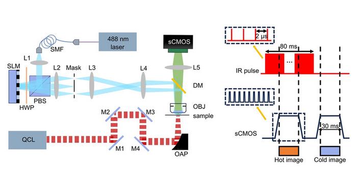
At the heart of the SIMIP system lies an innovative combination of a quantum cascade laser (QCL), which selectively excites molecular bonds and induces localized heating in the sample, and a structured illumination setup incorporating a continuous-wave laser and a spatial light modulator (SLM). The QCL is adept at inducing specific molecular vibrations, thereby resulting in minute temperature changes that can affect adjacent fluorescent molecules’ brightness. Concurrently, the structured illumination system projects intricate striped light patterns onto the sample from varying angles, creating Moiré fringes that encode previously unresolvable high-frequency details into lower-frequency signals. These modulated fluorescent signals are then captured by a scientific complementary metal-oxide-semiconductor (sCMOS) camera.
The process of capturing both the vibrational and fluorescent data enables SIMIP to reconstruct high-resolution images that are rich in chemical and spatial information, showcasing details that traditional methods might overlook. This reconstruction is achieved through sophisticated algorithms, namely Hessian SIM and sparse deconvolution techniques, pushing the boundaries of optical microscopy. Remarkably, the SIMIP technique has demonstrated spatial resolutions reaching up to approximately 60 nanometers, with an impressive imaging speed exceeding 24 frames per second, paving the way for a new generation of microscopic analysis.
One of the standout features of SIMIP is its ability to enhance the detection of autofluorescence, a natural phenomenon whereby certain biological molecules emit light without external labeling. By utilizing point-scanning SIM for structured excitation or shorter-wavelength probe beams alongside widefield photothermal detection methods, researchers can achieve greater compatibility with existing optical setups. This adaptability represents a significant advantage, allowing for broader applicability in varied research environments.
With its pioneering integration of structured illumination with mid-infrared photothermal imaging, SIMIP signifies a significant leap forward in our capacity for high-speed, super-resolution chemical imaging that transcends traditional diffraction limits. This novel approach ushers in exciting new opportunities within materials science, biomedical research, and chemical analysis, expanding the possibilities for detailed observations in the molecular landscape. The potential to utilize SIMIP for the detection of small-molecule metabolites and exploration of their interactions with cellular structures showcases its remarkable versatility and relevance to contemporary scientific inquiries.
Looking ahead, the research team envisions further refinements to SIMIP’s capabilities, including enhancements to its temporal synchronization aimed at increasing imaging speed and accuracy. They also plan to investigate the use of temperature-sensitive dyes to amplify sensitivity in various applications. These prospective developments, coupled with relatively minimal hardware adjustments to existing SIM systems, position SIMIP for swift adoption across laboratories worldwide, fostering a new era in microscopy.
The impact of this revolutionary technique is poised to resonate across diverse fields, from determining molecular interactions in complex biological systems to innovating imaging techniques within chemical analysis. As super-resolution imaging continues to evolve, tools like SIMIP will undoubtedly play a central role in unraveling the intricacies of the nanoscale world, granting researchers the unprecedented ability to observe, analyze, and understand the chemical and structural dynamics of matter at an extraordinary level of detail.
In conclusion, structured illumination mid-infrared photothermal microscopy represents a pivotal advancement in the realm of molecular imaging where high-resolution visualization intersects with rich chemical information. As researchers continue to unravel the complexities of this field, the intersection of technology and molecular science will undoubtedly lead to further breakthroughs, enhancing our understanding of the microscopic world and its implications for various scientific disciplines.
Subject of Research: VIRAL RESEARCH IN SUPER-RESOLUTION IMAGING
Article Title: Breaking the diffraction limit in molecular imaging by structured illumination mid-infrared photothermal microscopy
News Publication Date: 13-Apr-2025
Web References: SPIE Digital Library
References: 1. P. Fu, B. Chen, et al., doi 10.1117/1.AP.7.3.036003.
Image Credits: P. Fu, B. Chen, et al.
Keywords: Super-resolution microscopy, Structural illumination, Vibrational imaging, Chemical analysis, Nanoscale imaging, Quantum cascade laser, Autofluorescence detection, Photothermal microscopy.
Tags: challenges in vibrational imagingchemical contrast in microscopyenhancing spatial resolution in microscopyimaging without fluorescent tagsinnovative microscopy solutionsmid-infrared light absorptionmolecular identification techniquesnanoscale chemical imagingphotothermal microscopy advancementsstructured illumination microscopysuper-resolution microscopy techniquesvibrational imaging methods


