
In a significant advancement in the field of medical imaging, researchers from Kindai University in Japan have developed an innovative millimeter-wave sensor (MWS) designed to non-invasively monitor respiratory motion during crucial diagnostic procedures, such as X-ray and CT scans. This groundbreaking technology has the potential to transform how healthcare professionals manage and interpret images, addressing a longstanding challenge that has contributed to inaccuracies in diagnostic imaging.
Traditional methods of monitoring respiratory motion often rely on invasive or cumbersome technologies, such as infrared sensors that necessitate the use of reflective markers on a patient’s body. These approaches can not only compromise patient comfort but also result in inaccuracies due to the markers shifting or being misaligned during the imaging process. In stark contrast, the newly developed MWS operates without any direct contact, employing electromagnetic radiation to detect and visualize respiratory movements seamlessly, offering an unprecedented level of comfort and accuracy for patients undergoing imaging procedures.
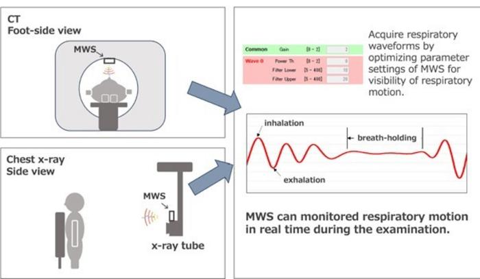
During diagnostic imaging, the ability to monitor respiratory motion accurately is critical to ensuring that images capture the required anatomical details without interference. Patients are often required to hold their breath to minimize motion artifacts in the resulting images, a task that can be challenging, particularly for children or patients with respiratory issues. The MWS fundamentally changes this dynamic by removing the need for reflective markers, thereby allowing patients to remain comfortable without the added pressure of maintaining a specific pose during imaging.
To validate the efficacy of the MWS, the researchers utilized a 24 GHz microMWS to assess its responsiveness to controlled respiratory motion. Through meticulous testing that involved a specialized breathing phantom, they were able to simulate different respiratory patterns consecutively. This method allowed for a robust comparison between the sensor’s detections and the established movements of the phantom, thereby substantiating the system’s reliability in diverse testing scenarios and confirming the sensor’s capability to accurately visualize subtle motion.
Moreover, a crucial aspect of this research involved extensive trials with an array of healthy volunteers, spanning a remarkable age range from six months to 64 years. This inclusive testing approach not only demonstrated the versatility of the MWS technology but also highlighted its capacity to adapt to a diverse patient population. Feedback from these trials reinforced the sensor’s non-contact advantages and its ability to capture vital respiratory data regardless of the subject’s clothing or positioning, whether supine or upright.
The implications of this research extend far beyond mere technical specifications; it strikes at the heart of improving patient experiences in diagnostic settings. The MWS system’s ability to deliver critical respiratory monitoring in real-time promises to elevate the standard of care delivered within hospitals and clinics. With tangible benefits such as reduced incidence of repeat imaging—often necessitated by poor image quality due to motion artifacts—the MWS could enhance workflow efficiency and ultimately safeguard patients’ exposure to unnecessary radiation.
The research team, comprising experts like Dr. Hiroyuki Kosaka, Dr. Kenji Matsumoto, and Dr. Hajime Monzen, believes that the MWS technology can set a new standard for respiratory monitoring across diagnostic imaging. By offering objective measurements that deliver immediate feedback, the potential exists to significantly decrease the number of repeat imaging sessions, which in turn can lead to faster diagnosis and improved treatment planning.
In addition to monitoring breathing movements accurately, the MWS technology is equipped with advanced capabilities to discern movement from various angles, further enhancing its usability in clinical settings. The researchers harnessed a radio-wave dark-box system to assess how effectively the sensor could capture motion while accounting for angle-related discrepancies. This capacity for multi-directional detection greatly amplifies the system’s clinical applicability in varying imaging environments.
Looking to the future, the researchers envision widespread integration of the MWS system into imaging protocols. Given its cost-effectiveness and user-friendly design, hospitals around the world could potentially deploy the device with relative ease, enhancing overall diagnostic accuracy and efficiency. Paramount among the benefits is the potential impact on vulnerable patient demographics, including elderly patients and children, for whom adhering to breath-holding protocols can prove particularly challenging.
The MWS technology presents an essential leap forward in how healthcare providers will approach respiratory motion monitoring in both imaging and radiation therapy. By providing a sophisticated, non-invasive option that improves diagnostic accuracy while conserving patient comfort, this development represents a significant breakthrough in medical technology. The ability to monitor respiratory movements with precision not only opens new avenues for clinical practice but ultimately fosters improved outcomes and experiences for patients.
The transformative nature of the MWS system may also lead to substantial advancements in research methodologies, prompting further investigations into respiratory dynamics during imaging procedures. As researchers continue to explore the complexities of respiratory motion and its impact on imaging quality, the insights gained from the MWS technology could stimulate innovation and guide the development of additional tools and techniques tailored to enhancing patient care across various medical specialties.
In conclusion, the advent of the millimeter-wave sensor heralds a new era of medical imaging, one in which the meticulously crafted union of technology, comfort, and accuracy comes to fruition. As the field of diagnostic imaging evolves alongside burgeoning technologies, the MWS stands ready to redefine standards and expectations, holding the promise of a brighter future for patients and healthcare providers alike.
Subject of Research: People
Article Title: Exploring the feasibility of millimeter-wave sensors for non-invasive respiratory motion visualization in diagnostic imaging and therapy
News Publication Date: 27-Jan-2025
Web References: DOI Link
References: None available
Image Credits: Dr. Hiroyuki Kosaka from Kindai University, Japan
Keywords
Health and medicine, Clinical medicine, Clinical imaging, Diagnostic imaging, Sensors
Tags: accuracy in diagnostic imagingadvancements in medical imagingchallenges in respiratory motion monitoringelectromagnetic radiation in healthcareinnovative imaging solutionsKindai University research breakthroughsmillimeter-wave sensor technologynon-contact respiratory motion monitoringnon-invasive medical technologiespatient comfort in diagnostic proceduresreducing motion artifacts in imagingX-ray and CT scan improvements