Drawing on studies of serum biomarkers, echocardiography, cardiac magnetic resonance imaging, nuclear medicine and clinical risk factors, the authors propose a three-stage framework combining early-warning screening, stratified evaluation and prognostic assessment. The review also stresses that polymyositis is now rarely diagnosed under modern classification criteria, so some findings apply to historical, mixed dermatomyositis/polymyositis cohorts whose members may now be classified differently. Idiopathic inflammatory myopathies include dermatomyositis, antisynthetase syndrome, immune-mediated necrotising myopathy, overlap myositis, inclusion-body myositis and polymyositis.
Both dermatomyositis and polymyositis characteristically cause symmetrical proximal muscle weakness, but the inflammatory process can also involve the myocardium, another type of striated muscle. Cardiac manifestations can include heart failure, myocarditis, arrhythmias and conduction abnormalities. Clinically overt involvement is relatively uncommon, yet cardiovascular disease remains an important cause of death.
Reported estimates of cardiac involvement range from 6% to 72%, while approximately 32.5% to 72% of patients have been reported to exhibit subclinical electrocardiographic abnormalities. Skin and skeletal-muscle manifestations can mask these cardiac signs, making early recognition difficult with conventional examinations. Population evidence indicates elevated cardiovascular risk among people with dermatomyositis or polymyositis, but the studies do not establish that inflammatory heart disease alone caused the difference.
A meta-analysis encompassing 22 studies found a pooled relative risk of 2.37 for cardiovascular events compared with the general population, with a 95% confidence interval of 1.86 to 3.02. Separate estimates were 2.35 for polymyositis and 2.55 for dermatomyositis, while ischemic heart disease had a relative risk of 1.76 and a 95% confidence interval of 1.40 to 2.21. In a study of 936 patients with dermatomyositis or polymyositis and 3,744 matched controls, the 10-year absolute risk of new-onset heart failure was 6.98%, compared with 4.58% in controls; atrial fibrillation risks were 10.17% and 7.07%, respectively.
Interpretation requires caution because matching on age and sex does not necessarily control for hypertension, diabetes, dyslipidaemia, obesity, smoking, older age or cumulative glucocorticoid exposure. The observed excess risk may therefore reflect an interaction among disease-related inflammation, treatment exposure and background cardiovascular risk. Biomarkers offer quantitative signals of myocardial injury, fibrosis and systemic inflammation, but their interpretation depends partly on their tissue origins and on what else is affecting a patient.
Cardiac troponin I is released exclusively by cardiomyocytes, whereas cardiac troponin T can also originate from injured adult skeletal muscle. In one inflammatory-myopathy study, cardiac involvement was present in 62% of patients with abnormal troponin I but 21% of those with abnormal troponin T, supporting greater cardiac specificity for troponin I. Creatine kinase was elevated in approximately 20% to 90% of dermatomyositis or polymyositis patients, sometimes reaching two to ten times the normal range, but it is not heart-specific. It can reflect skeletal-muscle injury and may rise during relapse, making it more informative about overall muscle disease than about the myocardium alone.
Myoglobin may change more rapidly after acute muscle injury, yet the review reports that it offers less value than creatine kinase in patients with chronic muscle damage. Emerging markers may reveal biological processes that precede visible cardiac dysfunction. Procollagen III N-terminal peptide and the carboxyterminal propeptide of type I procollagen are being investigated as indicators of active collagen synthesis and myocardial fibrosis.
In a study measuring serum PICP in 20 patients with dermatomyositis and 29 controls, the mean concentration was 326 plus or minus 76 ng/mL in patients and 135 plus or minus 88 ng/mL in controls, a difference with P less than 0.001; 60% of the patient group had elevated levels. Matrix metalloproteinases may reflect collagen degradation, and investigators have reported significantly elevated MMP-2 and MMP-9 messenger RNA in dermatomyositis/polymyositis muscle tissue, with no significant change in their tissue inhibitors. The microRNA hsa-miR-4442 was also higher in people with active dermatomyositis/polymyositis than in patients with other active autoimmune diseases or healthy controls.
Its current evidence mainly reflects overall disease activity rather than cardiac-specific involvement, so it remains exploratory rather than a validated marker of myocarditis or fibrosis. Autoantibodies can help identify patients who warrant closer cardiac evaluation, but the review states that current evidence does not support a validated antibody-based system for grading cardiac risk. Anti-Ro52 antibodies have been associated with glucocorticoid-resistant cardiac involvement and cardiac injury, although their specificity for cardiac disease is relatively low.
Anti-MDA5-positive dermatomyositis has carried an approximately 4% reported incidence of cardiac involvement, including new-onset atrial fibrillation or elevated cardiac troponin, while anti-Jo-1-positive juvenile dermatomyositis has been associated with more cardiovascular abnormalities. Anti-Mi-2-positive patients often have characteristic skin lesions and relatively little cardiac involvement, but a negative result does not exclude cardiac disease. Anti-SRP positivity is particularly associated with immune-mediated necrotising myopathy, a subtype that may include patients historically classified as polymyositis, and may identify a group with a higher prevalence of myocardial injury, arrhythmia and ventricular dysfunction.
These findings support antibody testing as part of a broader assessment rather than as a stand-alone cardiac test. Inflammatory markers provide another view of immune activity that may accompany cardiac inflammation. Interleukin-17 is implicated in the autoimmune inflammatory response and can increase REG3A in dermatomyositis/polymyositis, while REG3A has been studied as an indicator of cardiac inflammation and injury.
Tumour necrosis factor alpha is another central inflammatory cytokine; a polymorphism at TNF-alpha-308 has been associated with dermatomyositis susceptibility in one meta-analysis, while the A allele has been associated with ischemic heart disease risk in the general population in another. These findings make inflammatory markers potentially useful auxiliary warning indicators, but the review does not present them as a validated stand-alone cardiac risk-grading system. Multimodal imaging is central to detecting subclinical cardiac abnormalities that routine examination and conventional echocardiography may miss.
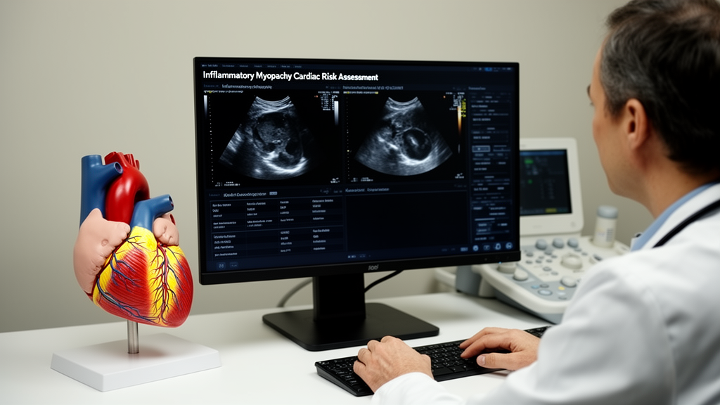
Echocardiography is non-invasive, convenient, repeatable and capable of real-time imaging, making it a practical first-line screening method. Tissue Doppler imaging captures myocardial motion signals and can supplement conventional measurements of structure and function. In a study of 30 patients with dermatomyositis or polymyositis who had no cardiovascular symptoms, conventional echocardiography combined with tissue Doppler imaging showed high sensitivity for identifying subclinical myocardial changes.
Speckle-tracking echocardiography assesses deformation through global longitudinal strain. Studies reviewed found reduced left- and right-ventricular global longitudinal strain in patients whose left-ventricular ejection fraction remained normal, indicating that subclinical systolic dysfunction can exist despite preserved conventional ejection fraction. Myocardial work is another proposed technique, combining strain with pressure estimates derived from a brachial-artery cuff to estimate contractile efficiency while reducing some afterload effects.
However, the review identified no direct studies applying myocardial work to dermatomyositis/polymyositis, so its value in this population remains prospective. Cardiac magnetic resonance imaging can detect and quantify myocardial oedema, fibrosis and inflammatory infiltration using T1- and T2-weighted imaging, native T1, extracellular volume and late gadolinium enhancement. It is also widely regarded as the reference standard for non-invasive quantification of ventricular volumes and ejection fraction.
A retrospective analysis of 44 dermatomyositis/polymyositis patients with normal left-ventricular morphology and function found elevated global native T1 and extracellular-volume values, suggesting that tissue characterization can detect early myocardial involvement before conventional dysfunction appears. Magnetic resonance imaging is more costly and time-consuming than echocardiography and is contraindicated for some patients with metallic implants, limiting its use as a universal screening test. Nuclear medicine methods provide complementary information about metabolism, perfusion and inflammation.
Technetium-pyrophosphate scintigraphy showed no significant difference in myocardial uptake between the reviewed patient cohort and healthy controls, although higher uptake correlated with left-ventricular diastolic dysfunction within the patient group. Fluorodeoxyglucose positron-emission tomography may assist with identifying myocarditis, but nuclear imaging is generally supplementary and often requires other tests and biomarkers for differential diagnosis. Based on the available evidence, the authors describe a proposed pathway beginning with clinical evaluation, cardiac troponin I, creatine kinase, inflammatory markers, myositis autoantibodies and conventional echocardiography.
Abnormal results can prompt electrocardiography, repeat echocardiography and, when appropriate, cardiac magnetic resonance imaging. Biomarker trends can then be interpreted alongside imaging changes to monitor cardiac function, but this strategy has not been directly validated prospectively in dermatomyositis/polymyositis. Endomyocardial biopsy remains the pathological reference standard for myocarditis, yet its invasiveness, sampling limitations and procedural risk restrict routine use.
It may be considered in selected cases of unexplained new-onset heart failure, refractory or fulminant arrhythmias, rapidly progressive cardiomyopathy or suspected myocarditis when non-invasive assessment is inconclusive and histology would affect treatment.
Subject of Research: Cardiac biomarkers, imaging and risk assessment in dermatomyositis and polymyositis
Article Title: Early warning and risk assessment of cardiac involvement in dermatomyositis/polymyositis: from biomarkers to multimodal imaging
Article References: Liu, W., Jiang, Y., Li, S., Song, C., Liu, K., Hou, M., Zhao, L., Li, W., Yan, B., & Mao, S. (2026). Early warning and risk assessment of cardiac involvement in dermatomyositis/polymyositis: from biomarkers to multimodal imaging. Frontiers in Medicine, 13, Article 1987083. https://doi.org/10.3389/fmed.2026.1987083
Image Credits: AI Generated
DOI: 10.3389/fmed.2026.1987083
Keywords: dermatomyositis, polymyositis, cardiac involvement, myocarditis, cardiac biomarkers, troponin, echocardiography, cardiac MRI, myocardial fibrosis, risk stratification, Early, warning
News Source: Ophelia Keating. (October 22, 2026). Review maps cardiac risk tools for inflammatory myopathies. Scienmag.