A new review published in Applied Microscopy provides a comprehensive guide for preparing forest pest specimens for X-ray micro-computed tomography, or XCT. The study, authored by Eun Jung Ahn and Ki Woo Kim, categorizes preparation methods into three distinct mounting types to optimize imaging quality for various insect life stages. This technique is increasingly vital for studying the internal structures of insects that threaten global forest ecosystems, offering a non-destructive alternative to traditional sectioning methods that only provide two-dimensional views. The review highlights that selecting the appropriate preparation method is essential for enhancing the quality of three-dimensional images, particularly given the varying sizes and types of forest pests.
X-ray micro-computed tomography is a powerful imaging technique that achieves micrometer-level resolutions, significantly higher than the millimeter-level resolutions typical of clinical computed tomography used in human medicine. Unlike traditional sectioning, which is limited to two-dimensional images, XCT allows for virtual sectioning that generates various types of three-dimensional images. This capability has made XCT widely used for histological analyses of animals and plants, enabling researchers to visualize complex internal structures without destroying the specimen. The application of XCT in insect research began in 2002, with early studies comparing it to traditional methods for analyzing insect heads, and it was first applied specifically to forest pests in 2017.
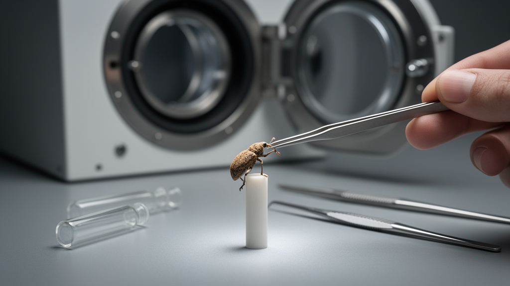
The review identifies three main categories of specimen preparation based on mounting types: simple mounting, liquid-cell mounting, and dry-cell mounting. Simple mounting is particularly suitable for adult beetles due to their hard exoskeletons, which provide natural structural support. In this method, specimens are often fixed, dehydrated, stained, and dried before being mounted on a holder, such as the tip of a fishing line. This approach minimizes specimen movement during scanning, ensuring better imaging quality.
Liquid-cell mounting is designed to preserve the soft tissues of larvae and pupae, which are more susceptible to damage during drying. In this method, the specimen is immersed in a liquid medium for scanning, effectively maintaining its structural integrity. This technique is particularly useful for analyzing the development of internal organs in immature stages of insects. For example, in studies of the Euwallacea validus beetle, liquid-cell mounting allowed researchers to examine the development of mycangia, specialized organs for carrying symbiotic fungi, in male and female larvae, pupae, and adults. The method involves fixing specimens in preservation liquid and scanning them while still submerged, facilitating detailed analysis of soft tissues that would otherwise be difficult to image.
Dry-cell mounting is used for immobilized live or freshly collected specimens that require anesthesia or euthanasia. It is particularly useful for studying live insects, such as the Cacosceles newmannii beetle, where repeated scans of live larvae were conducted to visualize and quantify their tracheal systems. In this case, larvae were anesthetized with sevoflurane and mounted in a closed-cell foam, allowing for detailed three-dimensional reconstruction of their internal structures.
The review discusses six representative cases to illustrate the application of these preparation methods. One case involves the coffee borer beetle, Hypothenemus hampei, which is the most significant pest of coffee trees. This beetle is only 1–2 mm in length, making it one of the smallest insects that can be analyzed by XCT. In one study, adult male and female beetles were fixed in ethanol, dehydrated, stained with iodine, and dried using hexamethyldisilazane. The resulting three-dimensional images revealed detailed anatomical differences between sexes, including the presence or absence of flight muscles and variations in the size and shape of the brain and midgut. The tracheal system was also observed to have a diameter narrower than one micrometer, highlighting the high resolution achievable with this technique.
Another case involves ambrosia beetles, which are wood-boring beetles that cultivate fungi for food. A study compared three visualization methods: paraffin sectioning, laser ablation tomography, and XCT. The specimens were cryopreserved in ethanol, dehydrated, and dried using a critical point drier or at room temperature. XCT was found to be the most efficient method for discovering new organs in ambrosia beetles, revealing different types of mycangia and associated muscles. This study demonstrated the efficacy of XCT in analyzing the internal structures of these insects, providing detailed visualizations that were not possible with traditional methods.
The harlequin ladybird, Harmonia axyridis, is another subject of the review. This species, native to Asia, has been introduced to North America and Europe as a biological control agent for aphids. However, it has become a pest itself, feeding on non-target insects and plants. A study used liquid-cell mounting to generate three-dimensional models of the prepupa and pupa stages, investigating changes in internal organs during metamorphosis. The specimens were anesthetized with carbon dioxide, preserved in ethanol, and stained with Lugol’s solution. The resulting images showed major internal changes, such as the development of indirect flight muscles in the dorsal plate during the late prepupal stage.
The review also highlights the importance of contrast enhancement in XCT imaging. Soft tissues often yield low-contrast images, requiring staining with contrast agents such as osmium tetroxide, phosphotungstic acid, or iodine. Osmium tetroxide is commonly used for electron microscopy, while phosphotungstic acid and iodine are preferred for their low toxicity. The drying step, which removes liquid from specimens, can improve image quality by enhancing the contrast of soft tissues, but it can also cause structural damage. Therefore, the choice of drying method, such as using a critical point drier or hexamethyldisilazane, depends on the susceptibility of the specimen to deformation.
The review concludes that the choice of preparation method should be based on the specific characteristics of the specimens and the research objectives. Simple mounting is best for adult beetles with hard exoskeletons, liquid-cell mounting is suitable for soft tissues of larvae and pupae, and dry-cell mounting is ideal for immobilized live specimens. The authors suggest that embedding methods may be beneficial for specimens with significant soft tissues, and that the combined use of XCT with electron microscopy could be highly beneficial for future studies. Additionally, the potential of XCT for studying parasites associated with forest pests is noted, with examples of its use in investigating parasitoid wasps within fly pupae.
Forest pests pose serious risks to biodiversity, timber production, and overall forest health, with their impact exacerbated by climate change. Rising temperatures and altered humidity levels can increase the frequency and severity of pest outbreaks, leading to larger insect populations inhabiting higher latitudes.
Specimen preparation for XCT involves a sequence of steps designed to prevent structural changes, avoid movement during scanning, and enhance contrast. The five main steps are fixation, dehydration, staining, drying, and mounting, though certain steps may be omitted depending on the sample type. Fixatives such as formalin and ethanol are necessary because insect specimens are often wet and fresh. Formalin induces the crosslinking of nucleic acids and proteins, which means specimens preserved in formalin are not appropriate for DNA and protein assays. Ethanol, on the other hand, dehydrates proteins, causing shrinkage of specimens. Selecting an appropriate preservation method is critical based on the specimen type to balance structural integrity with downstream analytical needs.
Enhancing contrast remains a primary challenge in the XCT of biological specimens, as soft tissues tend to yield low-contrast images. To differentiate soft tissues from other organs, staining with a contrast agent is required. The most commonly used contrast agents include osmium tetroxide, phosphotungstic acid, and iodine. Osmium tetroxide can fix and stain unsaturated fatty acids and is commonly used for electron microscopy specimen preparation. Phosphotungstic acid and iodine-based contrast agents are often selected for their low toxicity. The drying step, which removes liquid from specimens, improves image quality by enhancing the contrast of soft tissues. However, this process can cause structural damage. A critical point drier or hexamethyldisilazane is commonly used for drying, similar to electron microscopy protocols. Specimens less susceptible to deformation, such as wood samples dried at room temperature, can be directly scanned without these specialized drying methods.
For in vivo XCT, the X-ray source and detector are rotated to adjust for possible specimen movements, whereas in vitro XCT only requires specimen rotation. Anesthesia is required to immobilize live samples for in vivo XCT because images are generated by stacking multiple two-dimensional images. For common laboratory animals such as mice, rats, and rabbits, isoflurane is used for anesthesia. For insects, carbon dioxide or sevoflurane is used. Selecting the most appropriate mounting method is also important, where a non- or low-attenuating material is preferred for the sample holder. In the case of the harlequin ladybird, 200-microliter pipette tips were used as sample holders, with the tops trimmed and sealed at high temperatures. These tips were filled with phosphate-buffered saline and sealed with parafilm to prevent drying during the scanning process.
The application of XCT in insect research has evolved since its start in 2002. Early studies compared XCT with traditional sectioning methods for studying the head of Priacma serrata, highlighting the convenience of XCT for analyzing musculature. In 2007, researchers visualized the neural network of the brain of Drosophila by scanning larvae and producing comprehensive three-dimensional images of their central nervous system. Following these advancements, laboratory XCT systems became more widely distributed, leading to an increase in morphological studies of insects. In 2017, XCT was first applied to study forest pests, specifically Coleoptera, by examining the head of an adult poplar leaf beetle. The scanned images provided detailed visualizations of their muscles and nervous systems, demonstrating the technique’s utility for detailed anatomical analysis of forest-dwelling insects.
Subject of Research: Specimen preparation methods for X-ray micro-computed tomography of forest pests
Article Title: Specimen preparation for X-ray micro-computed tomography of forest pests
Article References: Ahn, E. J., & Kim, K. W. (2025). Specimen preparation for X-ray micro-computed tomography of forest pests. Applied Microscopy, 55(1), Article 3. https://doi.org/10.1186/s42649-025-00108-4
Image Credits: AI Generated
DOI: 10.1186/s42649-025-00108-4
Keywords: X-ray micro-computed tomography, forest pests, specimen preparation, insect imaging, mounting methods, biological imaging, entomology, non-destructive imaging, Specimen, preparation, X-ray, micro-computed
News Source: Denise Maddox. (November 12, 2026). Review details specimen prep for forest pest CT scans. Scienmag.