WASHINGTON — Researchers have developed an enhanced version of optical coherence tomography (OCT) that can image biomedical samples at higher contrast and resolution over a wider 3D field of view than was previously possible. The new 3D microscope could be useful for biomedical research and eventually enable more accurate medical diagnostic imaging.
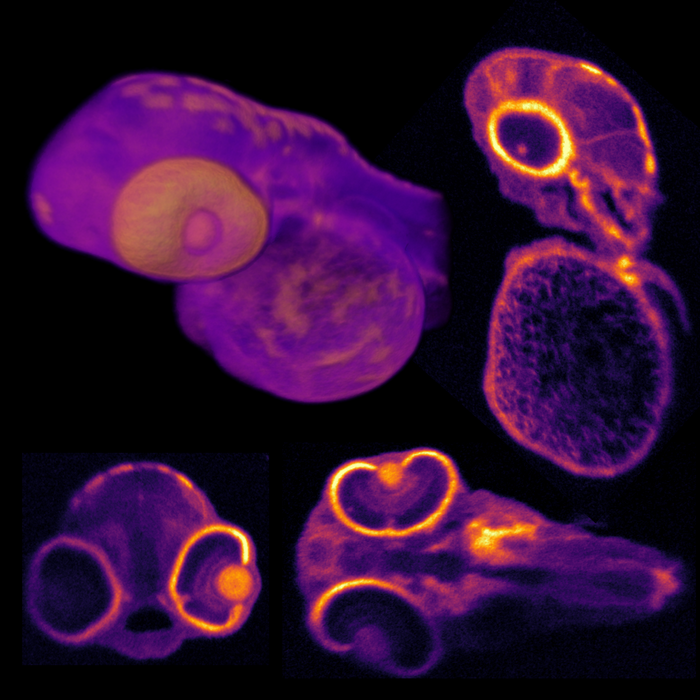
Credit: Kevin Zhou, Duke University
WASHINGTON — Researchers have developed an enhanced version of optical coherence tomography (OCT) that can image biomedical samples at higher contrast and resolution over a wider 3D field of view than was previously possible. The new 3D microscope could be useful for biomedical research and eventually enable more accurate medical diagnostic imaging.
In Optica, Optica Publishing Group’s journal for high-impact research, the researchers from Duke University describe the new technique, which they call 3D optical coherence refraction tomography (3D OCRT). Using various biological samples, they show that 3D OCRT produces highly detailed images that reveal features difficult to observe with traditional OCT.
OCT uses light to provide high-resolution 3D images without requiring any contrast agents or labels. Although it is commonly used for ophthalmology applications, the imaging method can also be used to image many other parts of the body such as the skin and inside the ears, mouth, arteries and gastrointestinal tract.
“OCT is a volumetric imaging technique widely used in ophthalmology and other branches of medicine,” said first author Kevin C. Zhou. “We developed a new and exciting extension, featuring novel hardware combined with a new computational 3D image reconstruction algorithm to address some well-known limitations of the imaging technique.”
“We envision this approach being applied in a wide variety of biomedical imaging applications, such as in vivo diagnostic imaging of the human eye or skin,” said research team co-leader Joseph A. Izatt. “The hardware we designed to perform the technique can also be readily miniaturized into small probes or endoscopes to access the gastrointestinal tract and other parts of the body.”
Seeing more with OCT
Although OCT has proven useful both in clinical applications and biomedical research, it is difficult to acquire high-resolution OCT images over a wide field of view in all directions simultaneously due to fundamental limitations imposed by optical beam propagation. Another challenge is that OCT images contain high levels of random noise, called speckle, which can obscure biomedically important details.
To address these limitations, the researchers used an optical design that incorporated a parabolic mirror. This type of mirror is commonly found in non-imaging applications, such as flashlights, where it surrounds the light bulb to direct the light in one direction. The researchers used an optical setup in which light was sent in the other direction, with the sample placed where the light bulb would be in a flashlight.
This design made it possible to image the sample from multiple views over a very wide range of angles. They developed a sophisticated algorithm to combine the views into a single high-quality 3D image that corrects for distortions, noise and other imperfections.
“The work published in Optica extends on our previous research by overcoming significant engineering challenges, both in the hardware and software, to allow OCRT to work in 3D and make it more widely applicable,” said research team co-leader Sina Farsiu. “Because our system generates tens to hundreds of gigabytes of data, we had to develop a new algorithm based on modern computational tools that have recently matured within the machine learning community.”
Getting a wider view
The researchers demonstrated the versatility and broad applicability of the method by using it to image various biological samples including a zebrafish and fruit fly, which are important model organisms for behavioral, developmental and neurobiological studies. They also imaged mouse tissue samples of the trachea and esophagus to demonstrate the potential for medical diagnostic imaging. With 3D OCRT, they acquired 3D fields of view of up to ±75° without moving the sample.
“In addition to reducing noise artifacts and correcting for sample-induced distortions, OCRT is inherently capable of computationally creating contrast from tissue properties that are less visible in traditional OCT,” said Zhou. “For example, we show that it is sensitive to oriented structures such as fiber-like tissue.”
The researchers are now exploring ways to shrink the system and make it faster for live imaging by taking advantage of recent developments in faster OCT system technologies and advances in deep learning that can speed up or improve data processing.
Paper: K. C. Zhou, R. P. McNabb, R. Qian, S. Degan, A.-H. Dhalla, S. Farsiu, J. A. Izatt, “Computational 3D microscopy with optical coherence refraction tomography,” 9, 6 (2022).
DOI: 10.1364/OPTICA.454860
About Optica
Optica is an open-access journal dedicated to the rapid dissemination of high-impact peer-reviewed research across the entire spectrum of optics and photonics. Published monthly by Optica Publishing Group, the Journal provides a forum for pioneering research to be swiftly accessed by the international community, whether that research is theoretical or experimental, fundamental or applied. Optica maintains a distinguished editorial board of more than 60 associate editors from around the world and is overseen by Editor-in-Chief Prem Kumar, Northwestern University, USA. For more information, visit Optica.
About Optica Publishing Group
Optica Publishing Group is a division of Optica, the society advancing optics and photonics worldwide. It publishes the largest collection of peer-reviewed content in optics and photonics, including 18 prestigious journals, the society’s flagship member magazine, and papers from more than 835 conferences, including 6,500+ associated videos. With over 400,000 journal articles, conference papers and videos to search, discover and access, Optica Publishing Group represents the full range of research in the field from around the globe.
Journal
Optica
DOI
10.1364/OPTICA.454860
Article Title
Computational 3D microscopy with optical coherence refraction tomography
Article Publication Date
2-Jun-2022


