A critical review of preclinical infectious disease modeling published in Laboratory Animal Research indicates that future investigations of emerging viral threats should transition away from using non-human primates as routine default screening platforms and instead implement a question-driven translational framework. Analyzing the preclinical research landscape reshaped by the coronavirus disease 2019 pandemic, researchers evaluated how non-human primate models contributed to deciphering severe acute respiratory syndrome coronavirus 2 infection dynamics and systemic host defenses. The analysis emphasizes that while non-human primates retain distinct utility when experimental questions require integrated analysis of multi-tissue pathology, systemic viral kinetics, longitudinal immune memory, and protective efficacy, their steep financial costs, constrained colony availability, specialized husbandry demands, and ethical considerations prevent routine utilization. Consequently, the authors propose a staged scientific workflow in which artificial intelligence, human-derived organoids, and microphysiological organ-on-chip platforms narrow evidence gaps and prioritize candidates upstream before conducting targeted, fit-for-purpose primate validation studies.
The biological rationale for deploying non-human primates during emerging viral outbreaks stems largely from their close phylogenetic proximity to humans, which is reflected in structural similarities between human and primate angiotensin-converting enzyme 2 receptors that facilitate productive viral entry and tissue tropism across Old World primate species. Furthermore, primate systems reproduce essential features of human disease pathology across multiple anatomical compartments and provide a mature immunological toolkit for evaluating protective immune durability and tissue-resident immunity. Controlled experimental environments allow researchers to perform defined inoculations, serial bronchoalveolar lavages, longitudinal thoracic imaging, and scheduled multi-tissue necropsies that would be impossible to conduct in clinical patient cohorts. Their comparatively long lifespans additionally enable direct tracking of durable immune memory and post-acute sequelae over extended intervals. These experimental capabilities established primates as indispensable tools during acute pandemic investigations, supporting evaluations of viral rechallenge, vaccine protection thresholds, and pharmacological interventions that guided clinical development programs.
Despite these experimental strengths, the authors caution that high internal validity achieved through tightly controlled animal cohorts can restrict external translational validity when findings are generalized to human populations. Laboratory studies typically employ synchronized exposures, uniform viral doses, defined inoculation routes, and genetically and environmentally homogeneous cohorts, which fail to replicate the complex exposure histories, circulating viral variants, diverse comorbidities, and heterogeneous immunological backgrounds found in clinical populations. In parallel, global policy shifts and primate infrastructure constraints are actively transforming the preclinical landscape. Regulatory initiatives such as the United States Food and Drug Administration roadmap for reducing animal testing through validated new approach methodologies, together with institutional transitions including the phase-out of monkey research at the Centers for Disease Control and Prevention and the Dutch decision to redirect Biomedical Primate Research Centre funding toward alternative methods by 2030, dictate that future infection research must employ primates more strategically and selectively rather than as routine initial screens.
Examining species-specific performance across macaque models reveals distinct experimental advantages and limitations that dictate their suitability for particular research questions. Rhesus macaques (Macaca mulatta) reproducibly develop substantial viral loads across both upper and lower respiratory tracts after intranasal and intratracheal challenge, manifesting interstitial pneumonia, pulmonary infiltrates, and transient clinical signs that generally resolve within one to three weeks. Supported by extensive immunological assays measuring binding antibodies, neutralizing titers, and functional T-cell responses, rhesus macaques represent the best-characterized primate model, though they predominantly exhibit mild-to-moderate illness rather than fatal respiratory distress. In contrast, cynomolgus macaques (Macaca fascicularis) also sustain productive viral replication but exhibit predominantly mild or asymptomatic clinical manifestations. However, inflammatory phenotypes and disease severity in cynomolgus macaques vary substantially with chronological age and host metabolic status, presenting opportunities to investigate host heterogeneity in structured cohorts, even though small sample sizes frequently limit statistical evaluations of inter-individual variation.
Other non-human primate species provide alternative phenotypes that address specific pathological and logistical questions outside the typical macaque spectrum. African green monkeys develop pronounced pulmonary inflammatory injury, particularly in aged animals, exhibiting diffuse alveolar damage, marked local interferon and interleukin-6 pathway activation, and progression toward acute respiratory distress syndrome-like pathology. Serial fluorine-18 fluorodeoxyglucose positron emission tomography combined with computed tomography facilitates sensitive non-invasive tracking of these inflammatory lesions in African green monkeys, even when overt clinical symptoms remain minimal. Baboons (Papio species), characterized by larger body sizes and naturally occurring age-related comorbidities in select colonies, display more prolonged pulmonary inflammatory responses than rhesus macaques, and their physical stature facilitates repeated vascular sampling and imaging-intensive longitudinal protocols, although direct evidence remains constrained by smaller historical datasets. Conversely, common marmosets (Callithrix jacchus) demonstrate low susceptibility, minimal viral replication, and only minor transient clinical changes, indicating that their restricted permissiveness makes them unsuitable as primary pathogenesis or efficacy models.
Evaluating vaccine efficacy and correlates of protection further demonstrates why preclinical animal models must be matched directly to explicit analytical objectives rather than chosen through a fixed hierarchy. Vaccine challenge investigations in rhesus macaques successfully defined quantitative thresholds linking prechallenge immune status to post-challenge protection, demonstrating that nucleoside-modified messenger RNA candidates elicit robust neutralizing antibody and T helper 1-biased cellular responses that substantially decrease viral loads in the lower respiratory tract. Passive antibody transfer experiments in rhesus cohorts confirmed dose-dependent protection mediated by immunoglobulin G, while revealing complementary protective roles played by cellular immunity when circulating antibody titers remain subprotective. Other species expand this analytical foundation, as cynomolgus studies evaluated immunogens against antigenically divergent strains, and baboon cohorts confirmed durable immunogenicity, wherein candidate vaccines elicited functional neutralizing antibody titers exceeding convalescent human plasma and maintained robust anamnestic responses upon booster administration one year later.
Therapeutic evaluations and immune memory investigations require similarly tailored model selections that reflect specific pharmacological mechanisms and anatomical compartments. Direct-acting antiviral agents, such as remdesivir, require models with consistent respiratory viral replication kinetics, and early intervention in rhesus macaques successfully demonstrated reductions in lung viral loads, pulmonary tissue pathology, and overall clinical severity. Conversely, host-directed or immunomodulatory therapies designed to curb excessive tissue damage demand pronounced inflammatory phenotypes, making African green monkeys biologically suited for anti-inflammatory testing, though published interventional studies in this model remain sparse. Regarding immunological memory, rhesus macaques established that primary exposure confers protection against rechallenge and drives systemic recall immunity against heterologous variants. Concurrently, cynomolgus macaque studies provided fine microanatomical resolution of tissue-resident immunity within draining lymph nodes and mucosal respiratory sites, revealing that viral clearance can occur without detectable neutralizing antibodies, while demonstrating that recall responses may not entirely avert transient immune-mediated pulmonary inflammation.
Investigating post-acute sequelae and extended longitudinal outcomes requires moving beyond acute viral clearance to measure protracted histopathological and extrapulmonary abnormalities. Longitudinal assessments in rhesus macaques detected persistent viral presence in tissues after the acute phase, alongside pulmonary accumulations of virus-associated foam cells displaying profibrotic and prothrombotic transcriptional signatures that point toward potential pathways for persistent tissue damage. In cynomolgus macaques, non-invasive whole-body immunoPET and nanobody-based positron emission tomography tracers enable serial visualization of widely distributed viral antigens and persistent systemic inflammatory changes without requiring premature terminal necropsies. African green monkeys expand longitudinal inquiries into systemic metabolic complications, exhibiting sustained hyperglycemia coupled with inflammatory signatures following viral challenge. While baboons offer physical dimensions suitable for extended serial imaging protocols, empirical data establishing their post-acute manifestations remain preliminary, underscoring that longitudinal programs benefit from merging the mechanistic depth of macaque necropsies with non-invasive imaging and extrapulmonary readouts in alternative primate species.
To streamline preclinical workflows and minimize unnecessary animal utilization, upstream computational platforms and artificial intelligence models are increasingly applied to resolve complex data spaces before launching animal experiments. Deep-learning and machine-learning frameworks integrate genomic sequences, structural biology parameters, and epidemiological surveillance records to predict viral mutational fitness, potential immune evasion, and emergent variant structures before specific strains achieve global dominance. Furthermore, artificial intelligence accelerates therapeutic development through automated drug repurposing, structure-guided antibody optimization, AlphaFold-assisted protein modeling, and multi-agent virtual screening workflows that drastically reduce empirical candidate libraries. Within active animal trials, computer vision algorithms and markerless pose estimation quantify subtle modifications in posture, mobility, social interactions, and home-cage behaviors, converting qualitative observations into objective, reproducible digital phenotypes. However, because computational algorithms remain vulnerable to incomplete training annotations, distribution shifts, and lack integrated physiological tissue contexts, their primary role is prioritizing hypotheses and candidates for experimental biological systems.
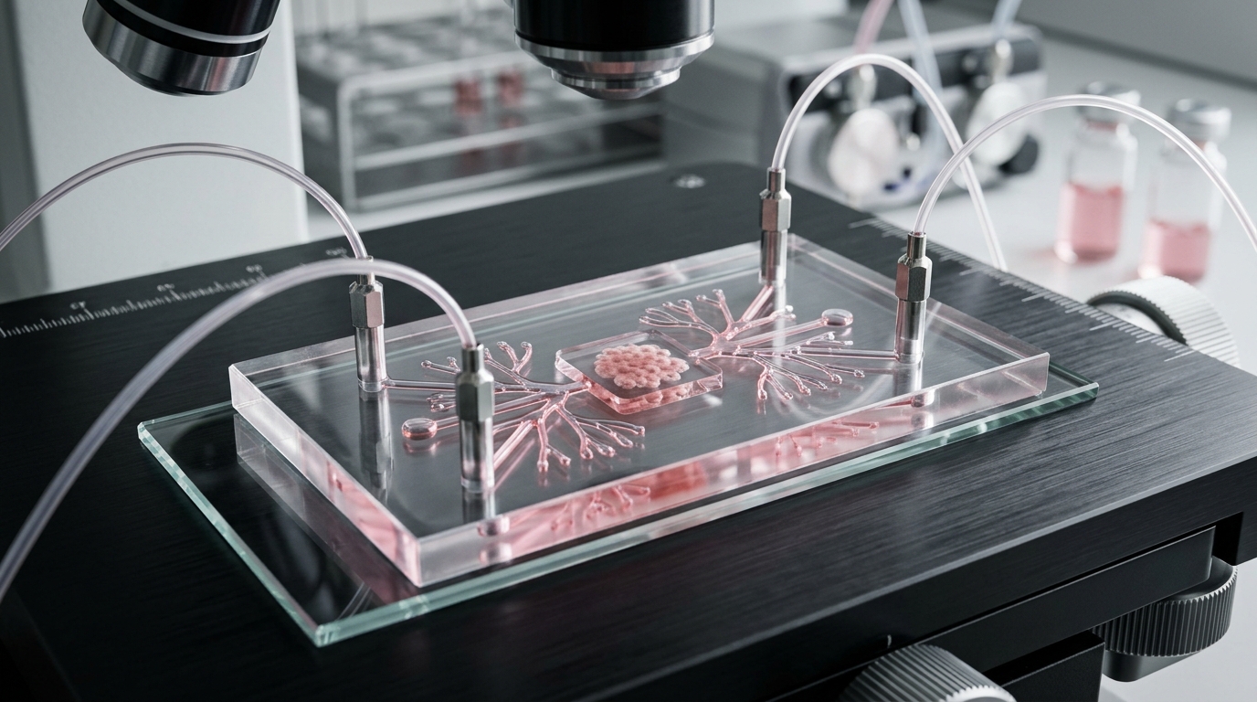
Human-derived organoid cultures and microphysiological organ-on-chip systems serve as intermediate mechanistic testing grounds that bridge computational predictions and whole-organism physiology. Three-dimensional organoids derived from human respiratory, intestinal, and neural tissues preserve primary cellular diversity and tissue architecture, allowing controlled characterizations of viral cellular tropism, epithelial barrier integrity, variant-specific replication kinetics, and innate host antiviral cascades. Microphysiological platforms augment static organoids by incorporating fluidic shear stress, tissue-tissue interfaces, and mechanical motion; for instance, lung-on-chip models simulate alveolar-capillary barrier degradation and immune-cell recruitment, while gut and blood-brain barrier chips capture enteric permeability, vascular dysfunction, and neuroinflammation. Complementary immune-mimetic constructs, such as the Modular Immune In vitro Construct and lymphoid-follicle chips, recreate human donor-derived immune cell interactions, cytokine release profiles, and vaccine-induced antibody production. Although these microscale systems currently lack full vascularization, mature adaptive immune circuits, and standardized inter-laboratory fabrication protocols, they reliably resolve human tissue-specific interactions that animal models cannot always replicate.
Synthesizing these complementary capabilities, the authors outline an integrated three-step translational pipeline that structures candidate progression through predefined go/no-go criteria. In this workflow, step one utilizes artificial intelligence and computational modeling to analyze genomic and epidemiological data, prioritizing high-fitness variants, optimal antigens, and lead therapeutic compounds. Step two deploys human organoids and organ-on-chip platforms to verify cellular infectivity, evaluate epithelial and vascular barrier integrity, and assess localized innate defenses under human-relevant conditions. Only candidates passing these upstream mechanistic filters advance to step three, where targeted non-human primate cohorts provide definitive whole-organism validation of systemic protection, complex inflammatory pathology, and durable immune memory. Discrepancies between computational forecasts, in vitro chip responses, and in vivo primate observations must be treated as critical decision-making checkpoints rather than experimental failures. By establishing rigorous, question-driven criteria that replace routine animal screening with fit-for-purpose primate validation, this staged framework strengthens experimental reproducibility, minimizes animal usage in alignment with 3Rs principles, and accelerates scientific preparedness for future emerging infectious diseases.
Subject of Research: Preclinical model selection and the integration of new approach methodologies with non-human primate studies in emerging viral infection research
Article Title: Rethinking non-human primate models for emerging viral infections after COVID-19
Article References: Kim, D.-Y., & Hong, J. J. (2026). Rethinking non-human primate models for emerging viral infections after COVID-19. Laboratory Animal Research, 42(1), Article 37. https://doi.org/10.1186/s42826-026-00299-1
Image Credits: AI Generated
DOI: 10.1186/s42826-026-00299-1
Keywords: SARS-CoV-2, non-human primates, rhesus macaque, cynomolgus macaque, African green monkey, baboon, marmoset, organoids, organ-on-chip, computational modeling, artificial intelligence, vaccine efficacy, viral pathogenesis, correlates of protection, post-acute sequelae, 3Rs principles, translational medicine
News Source: Kristina Jarvis. (November 17, 2026). Researchers Propose Staged Strategy Combining New Approach Methodologies With Primate Models. Scienmag.