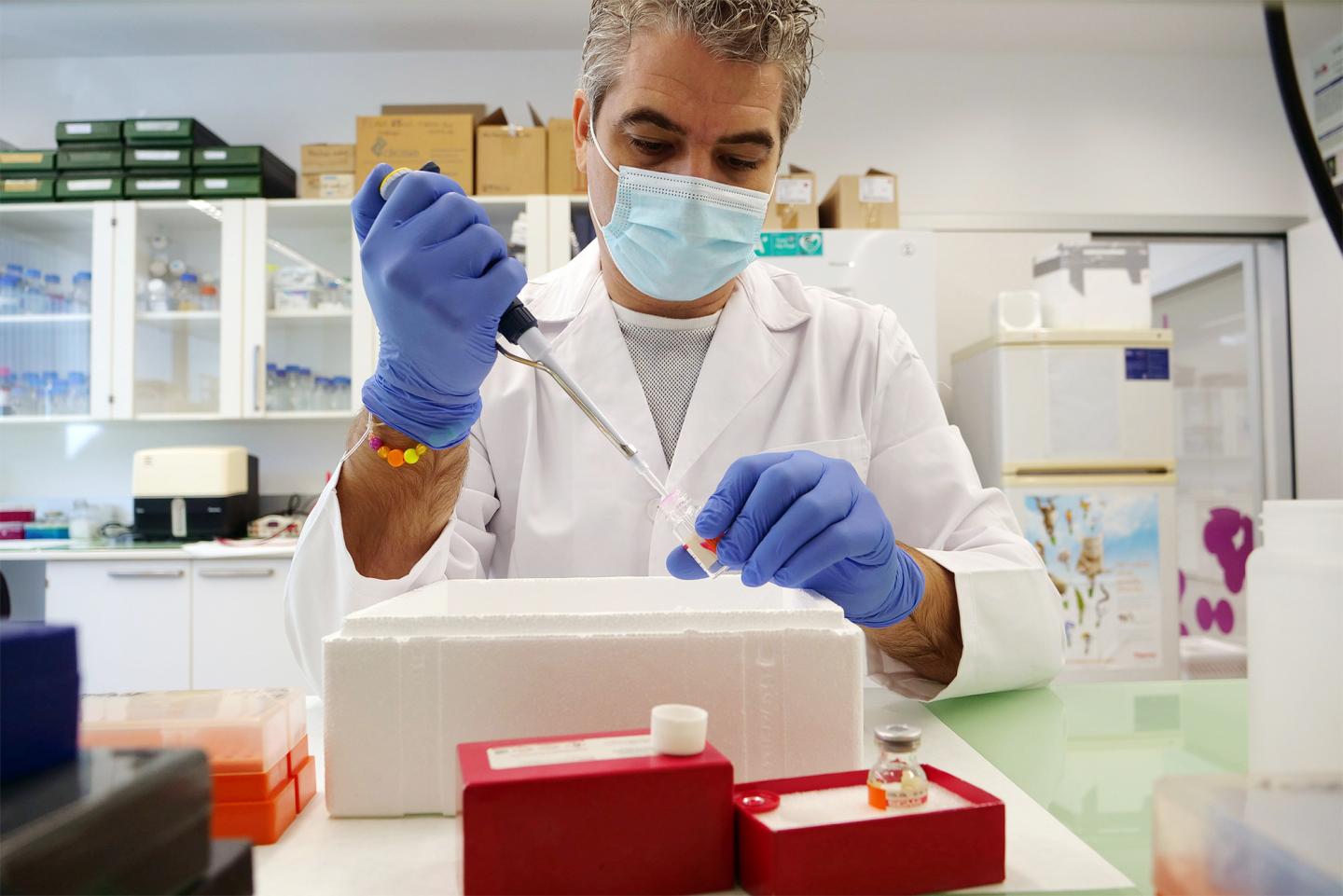
The researcher of the University of Malaga Adrián Ruiz-Villalba leads an international study that has revealed the molecular mechanisms involved in the activation of these cells

Credit: University of Malaga
The researcher of the Faculty of Science of the UMA Adrián Ruiz-Villalba, who is also member of the Andalusian Center for Nanomedicine and Biotechnology (BIONAND) and the Biomedical Research Institute of Malaga (IBIMA), is the first author of an international study that has identified the heart cells in charge of repairing the damage caused to this organ after infarction. This study has been recently published in the American Heart Association journal Circulation, first in the world dedicated to cardiovascular research.
These “reparative” cells consist in a population of cardiac fibroblasts that play a crucial role in generating the collagenous scar required to prevent ventricular wall rupture. This research, carried out with scientists from the Center for Applied Medical Research of Pamplona (CIMA) and the University Clinic of Navarra, also reveals the molecular mechanisms that activate these cells and regulate their function.
This discovery, according to the researchers, will enable the identification of new therapeutic targets and the development of targeted therapies to control the cardiac repair process following infarction.
Characterization of reparative cardiac fibroblasts
Cardiac fibroblasts are one of the essential components of the heart. These cells play a key role in maintaining the structure and mechanics of this vital organ. “Recent studies have shown that fibroblasts do not respond homogeneously to heart injury, so the purpose of our study was to define their heterogeneity during ventricular remodeling and understand the mechanisms that regulate the function of these cells”, explains Felipe Prósper, researcher of CIMA and the University Clinic of Navarra.
“Through single-cell transcriptomics (single-cell RNA-seq analysis) we identified a subpopulation within cardiac fibroblasts that we named Reparative Cardiac Fibroblasts (RCF) for their role after cardiac injury. We verified that, when a patient suffers a heart attack, these RCF activate to provide a fibrotic response that generates a collagenous scar that prevents heart tissue rupture”, says the researcher, who is also member of the Cell Therapy Network (TerCel) and the Health Research Institute of Navarra (IdiSNA).
CTHRC1, a protein related to the collagen that is essential for the healing process
At a molecular level, these scientists have discovered that RCF are characterized by a unique transcriptional profile, that is, a specific information pattern for the expression of the genes involved in their cardiac function. “Among the main differential markers of the transcriptome of these cells we identified the CTHRC1 protein (Collagen Triple Helix Repeat Containing 1), a molecule that is essential in the fibrotic response after myocardial infarction. Specifically, this protein participates in the collagen synthesis of the cardiac extracellular matrix and is crucial to the ventricular remodeling process”, says the researcher Adrián Ruiz-Villalba.
According to Ruiz-Villalba, these results suggest that RCF activate the cicatrization of cardiac injuries by segregating the CTHRC1 protein. Therefore, this molecule could be considered as a biomarker associated with the physiologic state of a damaged heart and a potential therapeutic target for patients that have had a myocardial infarction or that suffer from dilated cardiomyopathy.
This study falls within the line of research on cardiac fibrosis of the “Cardiovascular Development and Angiogenesis” group led by José María Pérez Pomares, professor of the Department of Animal Biology of the Faculty of Science of the UMA. This group studies the cellular and molecular mechanisms that underlie this type of pathologies by using different animal models and collaborating with different basic and clinical teams, both national and international.
‘Single-Cell RNA-seq Analysis Reveals a Crucial Role for Collagen Triple Helix Repeat Containing 1 (CTHRC1) Cardiac Fibroblasts after Myocardial Infarction’ is the result of five years of work, which comprises more than 30 different authors. Likewise, other R&D&I centers have participated in the study, such as NavarraBiomed (Pamplona), Maine Medical Center Research Institute (USA), CeMM Research Centre for Molecular Medicine of the Austrian Academy of Sciences (Vienna) and KU Leuven (Belgium).
###
Bibliography:
Ruiz-Villalba A. et al. Single-Cell RNA-seq Analysis Reveals a Crucial Role for Collagen Triple Helix Repeat Containing 1 (CTHRC1) Cardiac Fibroblasts after Myocardial Infarction. Circulation. 2020 Sep 25. Doi: 10.1161/CIRCULATIONAHA.119.044557
Media Contact
María Guerrero
[email protected]
Original Source
https:/
Related Journal Article
http://dx.


