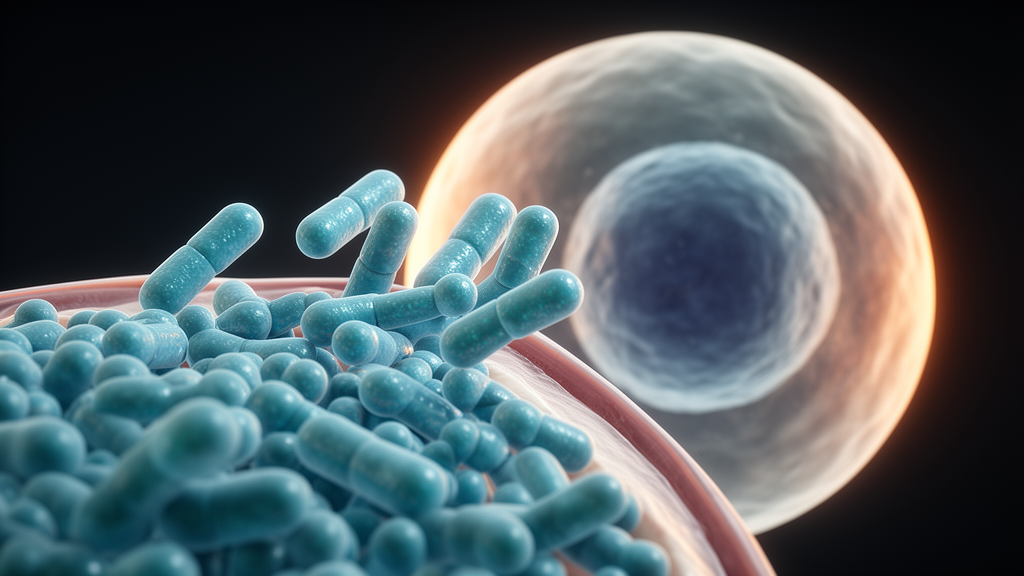
A class of synthetic polymers that mimic cell-penetrating peptides has shown a striking preference for acidic surroundings: in laboratory cultures of human cancer cell lines, the materials entered cells far more readily at mildly acidic pH than at the physiological pH of 7.4. The study, published as an open-access research article in Discover Polymers, evaluated so-called biodynamers—dynamic peptide analogs built from amino acid hydrazides and a carbazole dialdehyde monomer—as pH-responsive alternatives to conventional cell-penetrating peptides, which often lack targeting precision and raise toxicity concerns.
Biodynamers are assembled through constitutional dynamic chemistry. Amino acid hydrazides of lysine and arginine are linked to a hexaethylene glycol-conjugated carbazole dialdehyde through imine and acylhydrazone bonds, both of which are sensitive to acid. The resulting polymers fold in aqueous solution into amphiphilic nanorods roughly 10 nanometers long, with a hydrophobic carbazole core surrounded by a hydrophilic shell. The research team, led by Philipp Sonntag and Sangeun Lee of the Helmholtz Institute for Pharmaceutical Research Saarland collaboration network, synthesized three variants with different lysine-to-arginine ratios: Lys100-BD, Arg40Lys60-BD, and Arg80Lys20-BD.
Characterization confirmed the intended architecture. Size exclusion chromatography gave weight-average molecular weights of 6,090 daltons for Lys100-BD, 5,294 for Arg40Lys60-BD, 5,216 for Arg80Lys20-BD, and 5,062 for Arg100-BD, indicating that more arginine shortens the polymer. Dynamic light scattering yielded hydrodynamic diameters between 7.7 and 11.8 nanometers, consistent with earlier reports of rod-like structures confirmed by cryo-TEM. Electrophoretic light scattering showed that surface charge became more positive as arginine content rose, an expected consequence of arginine’s higher pKa and stronger protonation at neutral pH.
Before any uptake experiments, the team verified that the materials and the acidic test conditions themselves would not compromise the cells. In A549 lung adenocarcinoma and SW480 colorectal adenocarcinoma cells, none of the three biodynamers significantly reduced metabolic activity in MTT assays or damaged membranes in LDH assays across a concentration range of 16 to 256 micrograms per milliliter over 24 hours. The researchers also screened buffer systems: phosphate-buffered saline lacking calcium and magnesium dropped viability below the 80 percent threshold at pH 6.5, so pH-adjusted Hank’s balanced salt solution was selected for the pH experiments, and LDH testing confirmed membrane integrity at pH 7.4, 6.5, and 6.0 in both cell lines.
For the uptake studies, the biodynamers were labeled with rhodamine through their lysine side chains—a strategy that excluded the pure arginine variant, since NHS-coupling requires primary amines, and which the authors note may complicate intensity-based comparisons across compositions. Confocal microscopy and a cell lysis assay then quantified internalization after four hours in pH-adjusted HBSS. All three biodynamers showed significantly higher fluorescence signal at pH 6.0 than at pH 7.4 in both cell lines, measured by two-way ANOVA.
The magnitude of the effect depended on composition. In A549 cells, fluorescence-based quantification showed uptake increases of 5.3-fold for Lys100-BD, 4.6-fold for Arg40Lys60-BD, and 2.5-fold for Arg80Lys20-BD at pH 6.0 relative to pH 7.4. Under acidic conditions, Arg80Lys20-BD outperformed Lys100-BD by 3.7-fold and Arg40Lys60-BD by 2.6-fold, and the lysis assay confirmed its significantly higher uptake compared with both. SW480 cells showed the same direction of effect, with Arg80Lys20-BD again the strongest performer at low pH, though the fold increases were smaller.
The authors attribute the acid-triggered uptake to partial degradation of the dynamic covalent backbone. When pH drops, cleavage of the imine and acylhydrazone bonds exposes additional terminal amino and hydrazide groups, raising the effective charge density even as molecular weight falls. SEC measurements after four hours in pH-adjusted HBSS showed molecular weight reductions of 4.4 to 18.0 percent when pH dropped from 7.4 to 6.0, and zeta-potential measurements in the same buffer showed surface charge increases of roughly 1.6- to 3.5-fold. Since arginine and lysine are already nearly fully protonated at pH 7.4, the authors argue this charge gain must come from newly exposed groups rather than simple protonation. Enhanced electrostatic attraction to the negatively charged cell membrane is the proposed consequence.
Conformation may also play a role. Circular dichroism spectroscopy of Lys100-BD in phosphate buffer showed a shift from predominantly beta-sheet-like folding at pH 7.4 toward alpha-helical features at pH 5.5, with regular alpha-helical content rising from 0.3 to 0.4 percent and distorted helical structures from 2.2 to 3.4 percent, while beta-sheet content declined. Alpha-helical structure is a recognized motif in many conventional cell-penetrating peptides. The authors caution, however, that the CONTIN-based analysis was developed for peptides, and the aromatic carbazole backbone interacts strongly with polarized light, so further structural analysis is needed before firm conclusions about secondary structure can be drawn.
A first probe of the entry mechanism pointed to an active process. When A549 cells were incubated at 4 degrees Celsius, which rigidifies the membrane and suppresses energy-dependent endocytosis, internalization of the biodynamers was negligible compared with 37 degrees Celsius, and SW480 cells showed the same trend. This suggests uptake proceeds mainly through an active pathway, though the specific endocytic route remains unidentified. The authors also note that previous studies reported endosomal escape of biodynamers via a proton sponge effect following degradation inside endosomes, and the new confocal images show fluorescence accumulating around the nucleus after uptake—hints, they stress, that require further investigation of the materials’ full intracellular fate.
The findings remain at an early, in vitro stage: two cancer cell lines, rhodamine-based labeling with acknowledged limitations, large error bars in some lysis-assay measurements, and no non-degradable or uncharged control polymers to isolate the contribution of charge density from that of dynamic structure. The authors outline next steps including refined synthesis, advanced labeling strategies covering arginine-rich species, inhibitor-based uptake studies, and extension to more complex tissue models and in vivo systems. Within those limits, the study establishes a working methodology for assessing pH-dependent uptake of dynamic polymers and supports the idea that acid-labile, compositionally tunable nanorods could one day serve as selective delivery vehicles for the acidic microenvironments of tumors or inflamed tissue.
Subject of Research: pH-responsive biodynamer nanorods as cell-penetrating drug delivery materials evaluated in cancer cell cultures
Article Title: Evaluation of dynamic peptide nanorods as a unique pH-dependent cell-penetrating agent
Article References: Sonntag, P., Kamal, M. A. M., Passi, C., Bassil, J., Loretz, B., Lehr, C.-M., & Lee, S. (2026). Evaluation of dynamic peptide nanorods as a unique pH-dependent cell-penetrating agent. Discover Polymers, 3(1), Article 18. https://doi.org/10.1007/s44347-026-00046-8
Image Credits: AI Generated
DOI: 10.1007/s44347-026-00046-8
Keywords: biodynamers, cell-penetrating peptides, pH-responsive polymers, dynamic covalent chemistry, drug delivery, nanorods, arginine, lysine, A549 cells, SW480 cells, endocytosis, circular dichroism
News Source: Bethany Barker. (October 29, 2026). Research report: Evaluation of dynamic peptide nanorods as a unique pH-dependent cell-penetrating agent. Scienmag.