


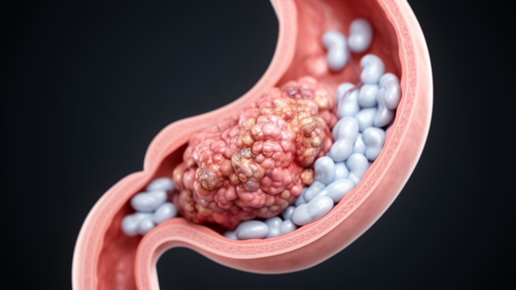
The report, published in the journal General Thoracic and Cardiovascular Surgery Cases, describes an esophageal adenosquamous carcinoma, a rare tumor type that contains both squamous cell carcinoma and adenocarcinoma components, whose lymph node metastases were overwhelmingly composed of mucinous carcinoma, the mucus-producing form of the adenocarcinoma.
Esophageal adenosquamous carcinoma accounts for roughly 0.2 to 1.0 percent of all esophageal malignancies. Its defining feature is the coexistence of two distinct cancer lineages within one tumor: a squamous component, which typically forms the superficial layer, and an adenocarcinoma component buried in the deeper tissue. That architecture creates a persistent diagnostic blind spot. Because endoscopic biopsy forceps sample only the surface, the deep adenocarcinoma component is frequently missed, and patients are often misdiagnosed before surgery as having the far more common esophageal squamous cell carcinoma. The optimal treatment strategy for the rare subtype remains unclear, and no standard chemotherapy regimen has been established for it.
The patient in this case arrived with dysphagia, a significant smoking history, and a medical history that included diabetes mellitus, myocardial infarction, prostate cancer, and bladder cancer. Laboratory testing showed an elevated carcinoembryonic antigen level of 13.6 nanograms per milliliter. Barium esophagography revealed an ulcerative lesion extending from the upper to the middle thoracic esophagus, and esophagogastroduodenoscopy identified a type 3 advanced tumor located 25 to 30 centimeters from the incisors. Biopsies taken from two sites told an unusual story: the oral portion of the tumor yielded squamous cell carcinoma, while an elevated anal lesion contained both squamous cell carcinoma and a mucinous carcinoma component, raising suspicion of adenosquamous carcinoma before surgery.
Contrast-enhanced computed tomography showed thickening of the esophageal wall along with enlarged lymph nodes along the right recurrent laryngeal nerve and in the middle thoracic paraesophageal region, with no distant metastases. Positron emission tomography-computed tomography added a puzzling detail that would prove consequential: the primary tumor was intensely fluorodeoxyglucose-avid, with a maximum standardized uptake value of 11.6, yet the suspicious regional lymph nodes showed no abnormal uptake at all. The team diagnosed clinical stage III (cT3N2M0) esophageal cancer with suspected adenosquamous carcinoma. Because the patient’s history of myocardial infarction and poor cardiac function made neoadjuvant chemotherapy unsuitable, he went directly to surgery: thoracoscopic esophagectomy with laparoscopic gastric tube reconstruction, three-field lymph node dissection, and jejunostomy.
The resected specimen confirmed the diagnosis and revealed a striking internal geography. The superficial portion of the tumor consisted of moderately differentiated squamous cell carcinoma, while the deeper layers contained mucinous carcinoma with abundant mucin and signet-ring cells, with transitional features between the two components. Immunohistochemical staining, including p40 positivity in the squamous element, carcinoembryonic antigen positivity in the glandular element, and Alcian blue staining of abundant mucin, confirmed squamous, glandular, and mucinous components. Notably, the elevated lesion that had yielded the mucinous component on biopsy corresponded not to the primary tumor’s own surface but to a metastatic lymph node that had directly invaded the esophageal wall, exposing the adenocarcinoma component to the luminal surface. That accidental exposure is what allowed both components to be captured preoperatively, something the authors note almost never happens.
The lymph node findings were the report’s most remarkable feature. Eighteen of 46 dissected lymph nodes were metastatic, classified as pN3, and involved the cervical, thoracic, and abdominal fields. In most involved stations the metastatic tissue was purely or predominantly mucinous carcinoma, accounting for approximately 60 to 100 percent of the deposit. A squamous cell carcinoma component appeared in only one station, the main bronchus node, where it made up about 25 percent of that deposit, and one small abdominal node near the lesser curvature contained pure tubular adenocarcinoma. Lymphatic and vascular invasion was almost entirely composed of the mucinous carcinoma component; although the squamous component was capable of nodal metastasis, no definite vascular invasion by the squamous element was identified. The final pathological stage was IVA (pT3N3M0).
This distribution inverts the pattern described in the prior literature. In previously reported esophageal adenosquamous carcinomas, nodal metastases predominantly comprised the squamous component, at rates of 60.9 to 85.7 percent, while the adenocarcinoma component alone accounted for only 0 to 8.7 percent. Against that benchmark, the mucinous carcinoma-predominant metastases in this patient are exceptional. The authors argue that this dissociation between a squamous-predominant primary lesion and mucinous-predominant metastases supports the view that the adenocarcinoma component governs the aggressive metastatic behavior of esophageal adenosquamous carcinoma, a hypothesis they frame cautiously as suggested by this single case rather than proven.
The mucinous predominance also retroactively explains the imaging anomaly. The authors conclude that PET-CT may underestimate nodal metastases in mucinous carcinoma-predominant esophageal adenosquamous carcinoma and that this limitation should be weighed during preoperative staging. Mucinous adenocarcinoma is itself rare among esophageal cancers, and mucin production has been proposed to promote invasion and metastasis by reducing intercellular adhesion and increasing tumor cell mobility; mucinous carcinomas of the colorectum and stomach are similarly associated with advanced disease and lymph node spread.
To place the case in context, the authors reviewed individual case reports published on PubMed over the past two decades, excluding case series, database studies, and review articles. Four cases, including the present one, were identified, with variable tumor locations and macroscopic appearances. In all cases except this one, the preoperative diagnosis was squamous cell carcinoma. They also summarized three single-institution retrospective studies: preoperative diagnosis was correct in only three of 38 patients in one series and one of 43 in another, and reported five-year overall survival rates ranged from approximately 30 percent to 64 percent. Although esophageal adenosquamous carcinoma has historically been regarded as aggressive, the largest surgical series, which used propensity-score matching, found overall survival after esophagectomy comparable to that of esophageal squamous cell carcinoma, while other reports described less favorable outcomes. The authors conclude that prognosis is at least as unfavorable as squamous cell carcinoma and is strongly influenced by nodal status and completeness of resection rather than being uniformly poor.
The clinical trajectory underscored the disease’s severity. The patient’s performance status deteriorated after surgery, so adjuvant chemotherapy was withheld. At seven months postoperatively, his carcinoembryonic antigen level rose and mediastinal lymph node recurrence was detected. With his general condition worsening, systemic chemotherapy remained contraindicated, and he died 11 months after the operation. Because he received no systemic therapy, the authors caution that no conclusions about chemotherapy sensitivity can be drawn from the case. They note that mucinous carcinomas in other organs are often less responsive to conventional chemotherapy, possibly because mucin impairs drug delivery, and suggest that biomarker evaluation and regimens used for esophagogastric adenocarcinoma may be worth considering in selected cases. Treatment of esophageal adenosquamous carcinoma generally follows surgery-based multidisciplinary principles developed for squamous cell carcinoma, including immune checkpoint inhibitor-based regimens for advanced disease. The authors emphasize that further accumulation of cases with detailed treatment and biomarker data is required to define optimal therapy for this rare tumor, and that the differential diagnosis should include mucoepidermoid carcinoma, a distinct entity in the World Health Organization classification that this case was able to exclude because both squamous and gland-forming adenocarcinoma components were clearly identified.
Subject of Research: A case report of esophageal adenosquamous carcinoma with mucinous carcinoma-predominant lymph node metastases
Article Title: Esophageal adenosquamous carcinoma with mucinous carcinoma-predominant lymph node metastases: a case report
Article References: Maeda, N., Yamamoto, M., Nakashima, R., Tajima, K., Seki, T., Ogimi, M., Kanamori, K., Ninomiya, Y., Kazuno, A., Shoji, Y., Kajiwara, H., Mori, M., & Koyanagi, K. (2026). Esophageal adenosquamous carcinoma with mucinous carcinoma-predominant lymph node metastases: a case report. General Thoracic and Cardiovascular Surgery Cases, 5(1), Article 30. https://doi.org/10.1186/s44215-026-00269-y
Image Credits: AI Generated
DOI: 10.1186/s44215-026-00269-y
Keywords: esophageal cancer, adenosquamous carcinoma, mucinous carcinoma, lymph node metastasis, case report, esophagectomy, PET-CT, histopathology, signet-ring cells, tumor staging, adenocarcinoma, surgical oncology
News Source: Nathaniel Bowman. (October 29, 2026). Research report: Esophageal adenosquamous carcinoma with mucinous carcinoma-predominant lymph node metastases: a case report. Scienmag.



