


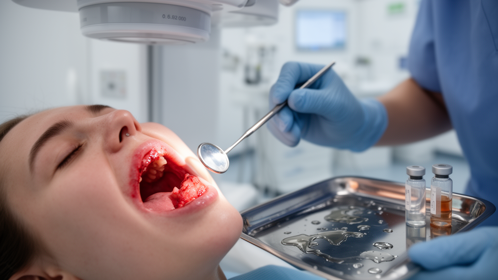
Patients with oral cavity cancer who undergo radiotherapy after surgery frequently develop oral complications, with mucositis affecting the great majority, but objective measures of salivary flow and gingival crevicular fluid volume showed no statistically significant overall change over the treatment period, according to a prospective study published in Supportive Care in Cancer. The findings, drawn from 52 enrolled patients of whom 28 completed both baseline and follow-up assessments, offer a detailed longitudinal picture of how the mouth responds during a treatment course that is known to challenge quality of life and treatment compliance.
Radiotherapy is a major treatment modality for head and neck cancers, and it can cause oral complications including mucositis, xerostomia, dysphagia and taste dysfunction. The study team, led by Mert Kutsal and colleagues, set out to evaluate longitudinal changes in salivary flow and gingival crevicular fluid volume and to explore associations between radiotherapy-related oral complications and periodontal status, radiation dose, volumetric parameters and tumor localization in patients with oral cavity cancer.
The researchers enrolled 52 patients with oral cavity cancer who had undergone surgery and were scheduled for radiotherapy. Clinical periodontal examinations and sampling of saliva and gingival crevicular fluid, the fluid that seeps into the small space between the tooth and the gum, were performed at baseline before treatment and again eight weeks after the start of radiotherapy. Radiotherapy-related oral complications were monitored throughout the study period. Because not every patient completed the second assessment, the team compared baseline demographic and clinical characteristics between those who did and did not complete follow-up to explore potential attrition-related differences, and found no statistically significant differences in any evaluated baseline characteristic, with all p values above 0.05.
Among the 28 patients with paired measurements, gingival crevicular fluid volumes averaged 50.80 microliters at baseline and 55.32 microliters at follow-up, while salivary flow rates averaged 0.34 milliliters per minute and 0.27 milliliters per minute, respectively. Neither gingival crevicular fluid volume, with a p value of 0.655, nor salivary flow rate, with a p value of 0.084, showed a statistically significant overall change across the treatment period.
An exploratory analysis stratified patients by prescribed primary planning target volume dose, comparing those prescribed 62 to 64 gray with those prescribed 66 to 70 gray. This stratification identified a significant time effect for salivary flow, with a p value of 0.042, whereas no significant change was observed in gingival crevicular fluid volume. A parallel stratification by elective planning target volume dose, comparing 54 to 56 gray with 58 to 60 gray, revealed no significant longitudinal changes in either gingival crevicular fluid volume, with a p value of 0.647, or salivary flow, with a p value of 0.089. The authors emphasize that these dose-stratified findings should be interpreted cautiously because of the limited analytical sample, substantial loss to follow-up, incomplete complication data and reliance on prescribed planning target volume doses rather than organ-at-risk dosimetry.
The oral complication analysis, which included the same 28 patients, found mucositis to be the most frequently observed complication, affecting 82.1 percent of patients, predominantly at Grade 1 to 2 severity. Xerostomia, the subjective sensation of dry mouth, followed at 39.3 percent, and dysphagia, difficulty swallowing, affected 35.7 percent.
The study’s statistical approach included descriptive, comparative and correlation analyses, with a p value below 0.05 considered statistically significant. The authors stress that the exploratory dose-stratified time effect for salivary flow should be viewed as hypothesis-generating rather than definitive, given the constraints of the dataset. They call for larger prospective studies with more complete follow-up and detailed dosimetric assessment to clarify the associations between radiation dose to specific organs at risk and the oral complications observed during treatment.
The authors’ conclusion that oral complications were frequently observed during radiotherapy in patients with oral cavity cancer stands alongside their caution that the limited analytical sample, substantial loss to follow-up and reliance on prescribed rather than measured doses temper the strength of any dose-response inference. The path forward, they write, lies in larger prospective studies with more complete follow-up and detailed organ-at-risk dosimetry.
Subject of Research: Longitudinal changes in salivary flow, gingival crevicular fluid and radiotherapy-related oral complications in patients with oral cavity cancer
Article Title: Evaluation of radiotherapy-related oral complications and functional outcomes in patients with oral cavity carcinoma
Article References: Kutsal, M., Akagunduz, O. O., Koca, K., Koska, S., Orman, M., & Becerik, S. (2026). Evaluation of radiotherapy-related oral complications and functional outcomes in patients with oral cavity carcinoma. Supportive Care in Cancer, 34(11), Article 1176. https://doi.org/10.1007/s00520-026-11413-3
Image Credits: AI Generated
DOI: 10.1007/s00520-026-11413-3
Keywords: oral cavity cancer, radiotherapy, mucositis, xerostomia, dysphagia, salivary flow, gingival crevicular fluid, periodontal status, radiation dose, supportive care, head and neck cancer, prospective study
News Source: Nathaniel Bowman. (October 30, 2026). Radiotherapy for oral cavity cancer: mucositis dominates while saliva and gum fluid stay largely stable. Scienmag.



