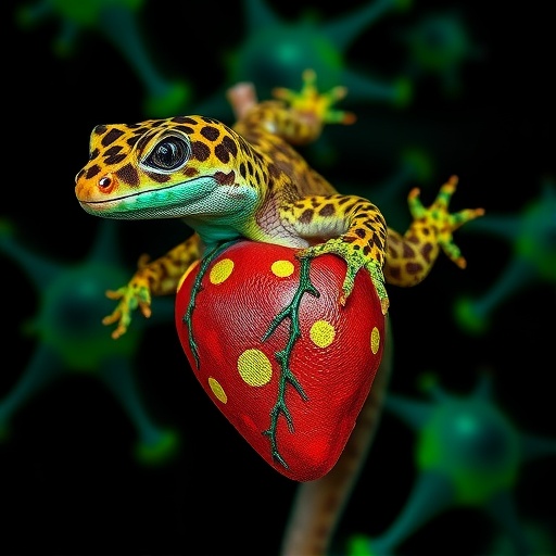
In a finding that could reshape how scientists think about repairing the damaged human heart, researchers at the University of Guelph have shown that the leopard gecko, a small terrestrial reptile best known as a popular pet, can regenerate its heart tissue after serious injury. Using a powerful technique known as quantitative proteomics, the team mapped, in unprecedented detail, the molecular choreography that unfolds inside the injured gecko heart over the course of one hundred days, and in doing so they identified a protein called agrin as a key player in the process. The work, published in BMC Genomics, represents the first time that an increase in agrin expression following cardiac injury has been reported in any species, and it positions the gecko as a valuable new model organism for studying cardiac repair.
The significance of the study lies in the kind of animal the researchers chose. Until now, cardiac regeneration has been characterized almost exclusively in fish and amphibians, such as zebrafish and salamanders, whose hearts are structurally simple and function at low blood pressures. Mammals, including humans, largely lose this ability shortly after birth; when our hearts are injured by a heart attack, the dead muscle is replaced by scar tissue rather than new functional muscle, setting the stage for heart failure. The leopard gecko, Eublepharis macularius, changes the calculus. Its heart is more complex than that of fish and amphibians, and it operates at higher pressures, making it anatomically and functionally closer to the mammalian heart. If a reptile with a comparatively sophisticated cardiovascular system can rebuild its own cardiac muscle, the molecular rules it follows may be more transferable to human medicine than those gleaned from fish swimming in cool, low-pressure aquatic environments.
To trigger regeneration, the team injured the gecko hearts using a cryoprobe, a chilled instrument that freezes and kills a small patch of cardiac tissue in a controlled and reproducible manner. This cryoinjury model closely mimics the kind of cell death seen in a human myocardial infarction. The researchers then collected tissue samples from the wound sites at four carefully chosen time points: three, fourteen, thirty, and one hundred days after injury. They also sampled the hearts of sham-operated geckos, animals that underwent the surgical procedure without actual cryoinjury, to serve as controls. By comparing the wounded hearts against these controls at each stage, the researchers could distinguish genuine injury responses from the nonspecific effects of surgery itself.
At the heart of the study’s methodology is quantitative proteomics, the large-scale measurement of proteins within a biological sample. While genomics tells researchers which genes are present or transcribed, proteomics reveals which proteins are actually being produced and at what abundance, offering a far more direct readout of cellular behavior. The team used high-resolution mass spectrometry to identify and quantify the proteins present in each sample, applying advanced bioinformatic tools to detect proteins whose levels changed significantly over the recovery period. They compiled and validated their results using specialized databases, including one built specifically for the leopard gecko proteome, and then performed Gene Ontology and pathway analyses to translate long lists of protein names into a coherent biological narrative about what the injured heart was doing at each stage of repair.
The results were striking. Across the time course, the researchers found that 579 proteins were differentially expressed at two or more time points, a substantial molecular signature of an active and dynamic repair process. Among the most important discoveries was a rise in the abundance of agrin at fourteen days post injury. Agrin is a protein best known for its role at the neuromuscular junction, but recent work in other contexts has shown that it can facilitate cardiomyocyte dedifferentiation, the process by which mature heart muscle cells essentially revert to a more primitive, proliferative state, allowing them to divide and produce new muscle cells. The gecko heart’s decision to ramp up agrin production precisely at the midpoint of its repair program suggests that dedifferentiation of existing heart muscle cells is a central mechanism of the regenerative response. This is the first report of injury-induced agrin upregulation in cardiac tissue, a finding that immediately suggests new avenues for therapeutic exploration in mammalian systems.
The Gene Ontology analysis painted a vivid picture of the metabolic and structural remodeling that accompanies regeneration. By fourteen days after injury, the wound site showed a coordinated decrease in oxidative phosphorylation and glycolytic capacity, indicating that the injured tissue had temporarily dialed down its energy production machinery. At the same time, the proteomic signatures of sarcomere organization, the intricate array of protein filaments that gives heart muscle cells their contractile power, and of mitochondrial content were markedly reduced. In essence, the cells at the injury site appeared to be dismantling their mature, specialized equipment, a hallmark of dedifferentiation. Heat shock proteins, molecular chaperones that stabilize and refold damaged proteins, were also modulated during this window, consistent with a tissue under acute stress while simultaneously reprogramming itself for growth.
What happened next is what makes the gecko heart so remarkable. By thirty days after injury, the regenerative program was visibly advancing, and by one hundred days the researchers could detect no Gene Ontology terms that differed between the regenerated hearts and those of sham-operated controls. The sarcomere organization had been rebuilt, mitochondrial populations had been restored, and the metabolic machinery of oxidative phosphorylation and glycolysis had returned to its baseline state. In other words, the gecko heart did not simply patch over the wound with scar tissue; it reconstructed the cellular architecture of the injury site so completely that, at the level of the proteome, the repaired tissue was indistinguishable from tissue that had never been damaged at all. This return to a pre-injury molecular state is the defining feature of true regeneration, and it is precisely what the mammalian heart fails to achieve.
The study’s conclusions emphasize that gecko heart regeneration involves the coordinated reorganization of cellular pathways governing mitosis, energy production, and contractile function. Rather than a single magic-bullet factor, regeneration appears to be a carefully timed sequence in which mature cardiomyocytes dedifferentiate, proliferate, and then redifferentiate, with the cell’s metabolic identity shifting in parallel from an energy-consuming, growth-oriented state back to the highly oxidative phenotype of a working heart muscle cell. The temporal resolution of the study, sampling at multiple stages across one hundred days, allowed the team to observe this sequence as it unfolded, capturing both the dismantling phase in the first two weeks and the reconstruction phase in the weeks that followed.
For the field of regenerative medicine, the implications are considerable. If the pathways that the gecko heart uses, including agrin-driven dedifferentiation, transient metabolic downregulation, and subsequent mitochondrial biogenesis, can be understood in sufficient molecular detail, researchers may be able to coax the mammalian heart into reactivating vestiges of these same ancestral programs. The finding that agrin expression rises after cardiac injury in a regenerating species is particularly compelling because agrin has already attracted attention as a possible therapeutic candidate, and the gecko data provide fresh evidence that this pathway is deployed during natural regeneration in a vertebrate with a complex, high-pressure heart. The researchers also note that proteins such as fibronectin, a component of the extracellular matrix involved in wound healing, showed dynamic changes across the time course, underscoring the importance of the structural scaffold on which new tissue is built.
The work also elevates the leopard gecko itself as an experimental model. Reptiles have been largely overlooked in regeneration research compared with fish and amphibians, yet the gecko has already demonstrated the ability to regenerate other tissues, and this study confirms that its cardiac regenerative capacity is robust and mechanistically accessible. The authors, led by Reece R. B. Long and senior investigators Todd E. Gillis, Matt K. Vickaryous, and Jennifer Geddes-McAlister at the University of Guelph, made their extensive proteomic datasets openly available as supplementary files, allowing other laboratories to mine the data for additional regenerative signatures. The research was funded by the Natural Sciences and Engineering Research Council of Canada, and all experiments were conducted under approved animal care protocols in accordance with national guidelines.
There remain, of course, many questions to answer. The proteomic approach reveals which proteins change in abundance but not always how they are regulated or what happens at the level of individual cells, and future studies combining proteomics with single-cell transcriptomics, imaging, and functional assays will be needed to fully dissect the gecko’s repair program. Nevertheless, the core message of the study is clear and hopeful: a vertebrate with a structurally complex, high-pressure heart can rebuild that heart completely after injury, and the molecular blueprint it uses is now coming into focus. In the gecko’s quiet, methodical restoration of its own cardiac muscle, researchers may be glimpsing a lost capacity of our own biology, one that modern medicine might eventually learn to reactivate in the millions of patients whose hearts cannot heal themselves.
Subject of Research: Cardiac regeneration in the leopard gecko (Eublepharis macularius), analyzed through quantitative proteomics of the cellular response to cryoinjury over a 100-day recovery period.
Subject of Research: Biology
Article Title: Quantitative proteomic analysis of the cellular response during cardiac regeneration in the leopard gecko (Eublepharis macularius)
Article References: Long, R. R. B., Jacyniak, K., Williams, C. J. A., Shaftoe, J. B., Geddes-McAlister, J., Vickaryous, M. K., & Gillis, T. E. (2026). Quantitative proteomic analysis of the cellular response during cardiac regeneration in the leopard gecko (Eublepharis macularius). BMC Genomics. https://doi.org/10.1186/s12864-026-13322-5
Image Credits: AI Generated
DOI: 10.1186/s12864-026-13322-5
Keywords: Cardiac regeneration, Cryoinjury, Agrin, Cardiomyocyte dedifferentiation, Quantitative proteomics, Cellular response, Metabolic shift, Mitochondrial biogenesis, Cardiac repair, Leopard gecko
Cite Scienmag News
APA MLA Chicago
Kenneth Gardner. (September 4, 2026). Proteomics reveals cellular mechanisms driving heart regeneration in leopard geckos. Scienmag. https://scienmag.com/proteomics-reveals-cellular-mechanisms-driving-heart-regeneration-in-leopard-geckos/
Kenneth Gardner. “Proteomics reveals cellular mechanisms driving heart regeneration in leopard geckos.” Scienmag, 4 September 2026, https://scienmag.com/proteomics-reveals-cellular-mechanisms-driving-heart-regeneration-in-leopard-geckos/. Accessed 4 September 2026.
Kenneth Gardner. “Proteomics reveals cellular mechanisms driving heart regeneration in leopard geckos.” Scienmag. September 4, 2026. https://scienmag.com/proteomics-reveals-cellular-mechanisms-driving-heart-regeneration-in-leopard-geckos/
Copy citation Download RIS
Tags: advanced proteomics techniques in cardiologyagrin protein in cardiac repairagrin protein in cardiac tissueanimal models for heart regenerationcellular pathways in heart regenerationcellular response to heart injurycomparative cardiac regeneration studiesevolutionary differences in heart regenerationheart regenerationimplications for human heart diseaseleopard gecko cardiac repairmolecular mechanisms of heart healingmolecular mechanisms of heart regenerationmolecular pathways in tissue regenerationpotential insights for human heart repairprotein mapping in injured heartsproteomics in heart healingproteomics in heart regenerationquantitative proteomics in cardiac researchregenerative biology in reptilesreptilian models of tissue regeneration