


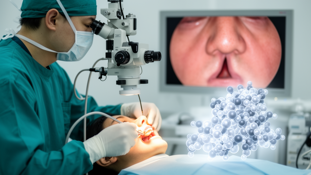
A protein that helps control how blood vessel cells multiply may hold clues to one of the most frustrating failures in eye surgery. The operation works well for most patients with chronic dacryocystitis, the tear-duct obstruction that causes constant watery eyes, sticky discharge and recurrent infection. Yet in roughly three percent of cases, the surgically created opening, called the ostium, scars over and closes again, and the tearing returns. A new study published in BMC Molecular and Cell Biology reports that patients whose openings re-blocked showed markedly higher levels of a protein called RBM39 in their nasal mucosal tissue, and that this protein drives proliferation of the endothelial cells that line blood vessels in laboratory experiments.
The research team, led by investigators at Tianjin Medical University Eye Hospital, collected nasal mucosal samples prospectively from 30 patients with chronic dacryocystitis undergoing En-DCR between February and June 2024. Fifteen of these patients later developed anastomotic re-obstruction within three months, confirmed by persistent tearing, visible scarring or granuloma blocking the ostium on endoscopic examination, and obstruction on lacrimal irrigation. The other fifteen, matched for age, sex and medical history, healed with a patent opening. The researchers emphasize that this is, to their knowledge, the first prospective proteomic analysis designed to identify molecular predictors of poor outcome after DCR surgery, comparing tissue taken at the time of the primary operation rather than after failure had already declared itself.
To profile the proteins in these tissues, the team used liquid chromatography coupled to tandem mass spectrometry, a technique that identifies and quantifies thousands of proteins simultaneously. From 3,056 detected proteins, they found 51 that differed significantly between the two groups when screened at a fold change of at least 1.5 and a p-value below 0.05: 26 upregulated and 25 downregulated. Among the proteins elevated in the re-obstruction group were SORBS2, SRSF9 and, most consequentially for this study, RBM39, an RNA-binding motif protein also known as HCC1 or CAPER alpha. Proteins reduced in the failing tissues included TMEM119, COL8A2 and several metabolic enzymes. The researchers chose RBM39 for deeper investigation because of its established links to microvascular density and endothelial expression, processes central to chronic inflammation and wound healing.
Validation experiments supported the proteomic signal. Immunohistochemistry on tissue sections showed that RBM39 was localized primarily to the vascular endothelium, the thin layer of cells lining blood vessels within the nasal mucosa. Western blotting, which measures specific proteins by antibody binding, confirmed that RBM39 levels were noticeably higher in tissue from the re-obstruction group than from patients with successful outcomes. Gene Ontology analysis of all 51 differentially expressed proteins pointed to enrichment at sites of focal adhesion and in RNA catabolism, while pathway analysis suggested involvement of the PPAR signaling pathway and RNA metabolism, hinting that both structural remodeling and post-transcriptional regulation accompany the failed healing response.
To test what RBM39 actually does, the researchers turned to HMEC-1, a well-characterized human microvascular endothelial cell line. Using small interfering RNA they knocked RBM39 down, and using lentiviral transduction they created cells that overexpressed it, verifying both manipulations at the mRNA and protein levels. Cell viability assays performed 72 hours after transfection showed that silencing RBM39 reduced proliferation, while forcing its expression increased it. Wound healing assays, which track how quickly a scratched cell layer closes, told the same story: knockdown impaired migration, overexpression enhanced it. Because endothelial proliferation and migration are the two cellular engines of angiogenesis, the results suggest RBM39 can push vessel-lining cells toward the behavior seen in exuberant granulation tissue.
The VEGF results added an intriguing asymmetry. Measuring vascular endothelial growth factor in the cell culture medium by ELISA, the team found that RBM39 overexpression significantly increased VEGF secretion, but knockdown produced no statistically significant decrease. The authors suggest that under low-expression conditions, other regulatory pathways may compensate to maintain basal VEGF, so the protein’s influence on this angiogenic factor only becomes visible when RBM39 is abundant. In a healing surgical wound, they argue, elevated RBM39 could tilt the local environment toward a pro-angiogenic state that favors the hyperplastic granulation tissue which serves as scaffolding for fibrotic scar.
Searching for the mechanism downstream, the researchers ran proteomic profiling on the manipulated cells. The knockdown comparison yielded 165 differentially expressed proteins and the overexpression comparison 409, and pathway enrichment analysis of the overexpression data highlighted spliceosomes, ribosomes, the HIF-1 signaling pathway and, most prominently, focal adhesion. Focal adhesions are macromolecular complexes that anchor the cell’s internal actin skeleton to the external matrix, and they function as hubs for adhesion, migration, proliferation and survival signaling. Western blotting confirmed that in RBM39-overexpressing cells, the focal adhesion proteins ACTN1 and, notably, VASP and Akt were shifted in ways consistent with the proteomic data, with ACTN1 downregulated and VASP and Akt upregulated.
The authors themselves are careful about how far these findings can be pushed. They note that no multiple-testing correction was applied to the differential protein screen, that differentially expressed proteins were selected on raw p-values and fold change without effect size evaluation, and that false positives from multiple comparisons are therefore possible. The tissue cohort was small, with only eight samples undergoing full proteomic analysis, and the findings require validation in larger independent cohorts. The functional work relied on a single endothelial cell line, whereas the real nasal mucosal wound involves a complex interplay of endothelial cells, fibroblasts, inflammatory cells and epithelial cells. Most importantly, the team states plainly that the current evidence remains primarily correlative: no pathway inhibition assays, transcriptional target validation or splicing analyses yet demonstrate that RBM39 directly controls ACTN1, VASP or the focal adhesion pathway, and no in vivo data exist on whether targeting RBM39 would actually prevent re-obstruction.
Even with those caveats, the study opens a concrete research direction. Existing work on postoperative nasal wound healing has concentrated on classic pro-fibrotic and pro-angiogenic mediators such as TGF-beta, VEGF and inflammatory cytokines, which the authors characterize largely as passive indicators of abnormal repair rather than upstream regulators. RBM39, by contrast, participates in alternative splicing and transcriptional co-regulation, positioning it as a potential upstream node that could coordinate endothelial behavior at both transcriptional and post-transcriptional levels. Anti-fibrotic agents such as mitomycin C have already been shown to improve success rates in endonasal DCR for patients with poor prognosis, so a molecular target identified prospectively from patient tissue could eventually complement such local interventions, though any therapeutic claim must await mechanistic and animal studies, which the authors propose to pursue in models such as rabbit lacrimal surgery.
For patients, the practical message today is modest but real: the molecular events that separate a healed tear duct from a re-blocked one are beginning to come into focus, and they involve far more than simple scarring. The prospective design means the tissue differences were present at surgery, before outcomes diverged, which is exactly what a predictive biomarker requires. If larger cohorts confirm that RBM39 levels at the time of En-DCR identify patients at risk of ostium re-obstruction, and if mechanistic work establishes how the protein drives endothelial proliferation through focal adhesion signaling, surgeons may one day have both an early warning sign and a pharmacological handle for the small but stubborn fraction of cases in which this elegant operation fails.
Subject of Research: Proteomic identification of RBM39 upregulation in nasal mucosal re-obstruction after endoscopic endonasal dacryocystorhinostomy
Article Title: RBM39 upregulation promotes ostium re-obstruction after endoscopic endonasal dacryocystorhinostomy (En-DCR)
Article References: Liu, J., Zhao, J., Zhu, X., Liu, X., Zhu, L., & Lin, T. (2026). RBM39 upregulation promotes ostium re-obstruction after endoscopic endonasal dacryocystorhinostomy (En-DCR). BMC Molecular and Cell Biology, 27(1), Article 33. https://doi.org/10.1186/s12860-026-00596-6
Image Credits: AI Generated
DOI: 10.1186/s12860-026-00596-6
Keywords: RBM39, dacryocystorhinostomy, proteomics, chronic dacryocystitis, vascular endothelial cells, focal adhesion pathway, VEGF, nasal mucosa, surgical failure, wound healing, HMEC-1, biomarker
News Source: Ophelia Keating. (October 29, 2026). Protein Linked to Re-Blocked Tear Ducts After Keyhole Eye Surgery. Scienmag.



