For decades, the identity of the cells that build and repair the lining of our blood vessels has been one of the most contested questions in vascular biology. Now, an international team led by researchers at The University of Queensland has brought unprecedented clarity to the field, showing that two seemingly unrelated molecular markers, when co-expressed, define a far more potent endothelial progenitor cell than any previously described. The work, published in the journal Angiogenesis, resolves longstanding contradictions between competing research camps and provides a refined, functionally validated definition of the cells responsible for vascular regeneration.
Since 1997, when Toshio Asahara and colleagues first reported the isolation of putative endothelial progenitor cells from human blood, researchers have argued about whether these progenitors arise from hematopoietic or mesenchymal lineages, whether they circulate through the bloodstream or reside permanently in vessel walls, and whether they are strictly unipotent or capable of producing multiple cell types. The confusion has been compounded by a proliferation of markers, each associated with some degree of regenerative capacity but rarely reconciled with the others. The new study set out to cut through this thicket by directly testing where candidate markers overlap within the endothelial hierarchy and which overlapping cells actually behave like true progenitors.
The researchers began with single-cell RNA sequencing of the mouse aorta, re-analyzing previously published data from the CD34-positive compartment at higher clustering resolution. This revealed distinct subclusters: differentiated endothelial cells expressing classic markers such as Pecam1 (CD31) and Cdh5 (VE-cadherin); mesenchymal clusters; and a transitional cluster, previously identified as the endovascular progenitor or EVP population, that expressed a mixture of both endothelial and mesenchymal genes. Among the candidate progenitor markers examined, only two stood out as enriched in this EVP cluster: Procr, encoding the Protein C Receptor, and Pdgfra, encoding Platelet-Derived Growth Factor Receptor Alpha. By contrast, CD157, ABCG2 and the transcription factor SOX18, all previously proposed as endothelial stem cell markers, were actually more highly expressed in differentiated endothelial cells than in the progenitor fraction, a finding that challenges the literature built around those molecules.
Flow cytometry on adult mouse aortae confirmed the transcriptomic picture at the protein level. Roughly 78 percent of EVPs expressed PROCR and about 82 percent expressed PDGFRA, and the two markers overlapped heavily within the same cells. Remarkably, when the researchers gated live aortic cells simply on the co-expression of PROCR and PDGFRA, more than 91 percent proved to be lineage-negative, VE-cadherin-positive endothelial cells, and nearly 95 percent of those fell into the CD34-positive, CD31-low EVP gate. In other words, two surface markers alone were sufficient to capture the progenitor population with around 95 percent accuracy, bypassing the complicated multi-parameter gating strategies previously required. The team named this double-positive population the refined endothelial progenitor cell, or rEPC, explicitly distinguishing it from the controversial circulating “EPCs” of the classical literature.
Functional testing was where the new definition earned its weight. When sorted cells were plated in limiting dilution colony-forming assays, rEPCs formed colonies in approximately 18 percent of wells, more than double the rate of PROCR-negative EVPs at 8 percent. Crucially, every rEPC colony displayed classical endothelial morphology and stained positive for Isolectin, an endothelial marker, whereas PROCR-negative EVP colonies were elongated and Isolectin-negative, and differentiated cells, regardless of PROCR status, formed no endothelial colonies at all. The team stresses that their rEPCs are non-hematopoietic, vessel-resident progenitors, a definition deliberately decoupled from the ambiguous circulating cells that dominated earlier decades of EPC research.
The most demanding test came in vivo. The investigators embedded 100 fluorescently labeled cells in collagen gels and implanted the gels under the skin of immunodeficient NSG mice. After seven days, gels seeded with rEPCs had engrafted in 9 of 15 cases, with an average of 5.7 percent of the plug area occupied by fluorescent, endothelial-marker-positive cells. Gels containing PROCR-negative EVPs engrafted in only 2 of 10 cases with roughly 0.8 percent coverage, and differentiated endothelial cells never engrafted, matching the acellular controls. Immunostaining of the retrieved gels showed that rEPC-derived cells co-expressed CD34, ERG and Isolectin, confirming they had generated genuine endothelial tissue within the host environment.
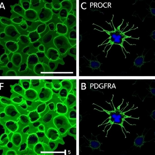
The study also mapped where these progenitors live. Immunofluorescence of aortic segments from reporter mice revealed that PROCR-positive endothelial cells cluster overwhelmingly in the intima of the thoracic aorta, where 68 percent of endothelial cells expressed PROCR, compared with just 21 percent in the abdominal segment. Function followed geography: endovascular progenitors sorted from the thoracic aorta formed endothelial colonies in 7 of 13 cultures, while abdominal counterparts formed none. The authors suggest this thoracic niche may reflect differences in embryonic origin, hemodynamic forces and local signaling environments that favor maintenance of a progenitor state.
Lineage tracing provided the final, formal proof that rEPCs actually generate new endothelium. Using Pdgfrα-MerCreMer/ROSA-EYFP mice, the team permanently labeled PDGFRA-expressing cells with yellow fluorescent protein and followed their fate. In the healthy, homeostatic aorta of juvenile mice, nearly all labeled endothelial cells were initially EVP-like progenitors rather than differentiated cells. By day 84, the proportion of labeled differentiated endothelial cells had risen significantly, demonstrating that labeled rEPCs had given rise to mature vessel-lining cells over time. The same tracing was applied to full-thickness skin wounds: at day one after injury, labeled cells in the wound center were exclusively progenitor-like, but by day five, labeled cells co-expressing CD31 and ERG appeared, showing active differentiation into mature endothelium at the site of tissue repair.
The findings translated to human tissue with notable consistency. Re-analysis of publicly available human normal aorta single-cell data identified clusters resembling the murine rEPC population, with CD34, PROCR and PDGFRA expression concentrated in these endothelial-mesenchymal transitional clusters. More strikingly, in endothelial colony-forming cells isolated from freshly donated human term placenta, adding a PROCR gate dramatically changed outcomes. CD31-intermediate cells that were PROCR-positive showed 90 percent colony-forming capacity compared with 15 percent for their PROCR-negative counterparts. Fully 80 percent of the PROCR-positive colonies reached high-proliferative-potential status, meaning they exceeded 1,000 cells and could form secondary colonies, and these cultures could be serially passaged to at least passage six while maintaining expression of CD31 and VE-cadherin. PROCR-negative colonies, in contrast, stalled at fewer than 50 cells and died. Human PDGFRA co-expression was supported only at the transcriptomic level, and the authors acknowledge that functional validation of double-positive human rEPCs remains an important goal for future work.
The implications extend across regenerative medicine, wound healing and tissue engineering. A short list of accessible surface markers that reliably predicts clonogenic, self-renewing, vessel-forming capacity could streamline the isolation of endothelial progenitors for therapeutic vascularization of engineered tissues, improve understanding of impaired wound repair in diabetes and aging, and inform studies of how tumors co-opt progenitor cells to build their blood supply. The study also delivers a cautionary message: several markers long used to isolate “endothelial stem cells,” including CD157, ABCG2 and SOX18, mark differentiated cells rather than progenitors in the aorta, suggesting that at least some published progenitor populations may need re-evaluation in light of functional rather than purely descriptive criteria. By fusing molecular definition with rigorous functional assays and lineage tracing, the work provides the vascular biology community with something it has lacked for nearly thirty years: a progenitor it can actually agree on.
Subject of Research: Functional definition and validation of refined endothelial progenitor cells (rEPCs) identified by PROCR and PDGFRA co-expression in mouse and human blood vessels
Subject of Research: Cancer
Article Title: Functional definition of endothelial progenitors by PROCR and PDGFRA co-expression
Article References: Styke, C., Kaur, S., Sim, S.-L., Zhao, J., Zhou, C., Wong, H. Y., Patel, J., Roy, E., Yoder, M. C., Harvey, R. P., Shafiee, A., & Khosrotehrani, K. (2026). Functional definition of endothelial progenitors by PROCR and PDGFRA co-expression. Angiogenesis, 29(4), Article 54. https://doi.org/10.1007/s10456-026-10078-0
Image Credits: AI Generated
DOI: 10.1007/s10456-026-10078-0
Keywords: endothelial progenitor cells, rEPC, PROCR, PDGFRA, endovascular progenitors, angiogenesis, vasculogenesis, lineage tracing, aorta, wound healing, endothelial colony-forming cells, vascular regeneration
Cite Scienmag News
APA MLA Chicago
Nathaniel Bowman. (September 7, 2026). PROCR and PDGFRA co-expression functionally identifies endothelial progenitor cells. Scienmag. https://scienmag.com/procr-and-pdgfra-co-expression-functionally-identifies-endothelial-progenitor-cells/
Nathaniel Bowman. “PROCR and PDGFRA co-expression functionally identifies endothelial progenitor cells.” Scienmag, 7 September 2026, https://scienmag.com/procr-and-pdgfra-co-expression-functionally-identifies-endothelial-progenitor-cells/. Accessed 7 September 2026.
Nathaniel Bowman. “PROCR and PDGFRA co-expression functionally identifies endothelial progenitor cells.” Scienmag. September 7, 2026. https://scienmag.com/procr-and-pdgfra-co-expression-functionally-identifies-endothelial-progenitor-cells/
Copy citation Download RIS
Tags: angiogenesis and vascular regenerationAngiogenesis journal publicationblood vessel lining repairblood vessel repair mechanismscell surface markers for vascular regenerationcell surface markers in vascular repairendothelial cell differentiationendothelial cell lineage identificationendothelial hierarchy and differentiationendothelial hierarchy and lineage tracingendothelial progenitor cell characterizationendothelial progenitor cell identificationhematopoietic vs mesenchymal originshematopoietic vs mesenchymal progenitorsmolecular markers for progenitor cellsmolecular markers in vascular biologyProcr and PDGFRA co-expression in endothelial progenitor cellsredefining endothelial progenitor cell identityvascular biology and regenerative medicinevascular biology research advancesvascular regeneration markers