In an illuminating development in cancer biology, researchers from the University of Eastern Finland have unveiled compelling insights into how pro-inflammatory M1 macrophages, critical players of the immune response, accelerate melanoma progression via the extracellular vesicles (EVs) they secrete. Published recently in the prestigious journal Cell Communication and Signaling, this study delineates a novel molecular crosstalk underlying the tumor microenvironment that potentiates melanoma aggressiveness through inflammatory mechanisms.
Macrophages are quintessential white blood cells tasked with immune defense, homeostasis, and tissue remodeling. Within tumors, these immune cells infiltrate and adopt diverse phenotypes broadly classified as pro-inflammatory M1 or anti-inflammatory M2 macrophages, influencing cancer fate dramatically. While M1 macrophages are traditionally linked to antitumor immunity, paradoxically, this study reveals that their secreted extracellular vesicles paradoxically foster melanoma cell invasion and inflammation, redefining their functional narrative in oncology.
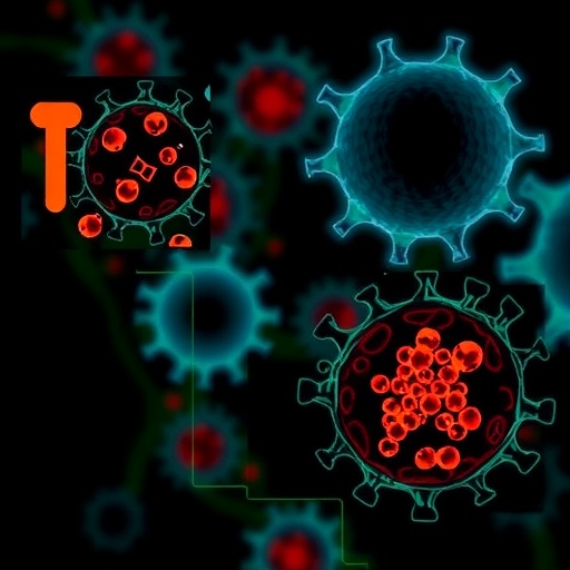
Extracellular vesicles are nano-sized, lipid bilayer-enclosed particles shed by virtually all cell types, carrying a rich cargo of proteins, lipids, and nucleic acids. These vesicles serve as sophisticated messengers facilitating intercellular communication, especially within the tumor microenvironment where they modulate cellular phenotypes, immune responses, and metastasis. The UEF research specifically analyzed EVs secreted by M1 macrophages and characterized their contents and effects on melanoma cells in vitro.
Advanced molecular assays identified critical pro-inflammatory cytokines, particularly tumor necrosis factor-alpha (TNFα) and interleukin-1 beta (IL-1β), encapsulated within the M1 macrophage-derived EVs. These potent signaling molecules are delivered directly into melanoma cells through vesicular internalization, orchestrating inflammatory signaling cascades intracellularly. The study revealed that the nuclear factor-kappa B (NF-κB) pathway, a master regulator of inflammation and immune responses, is robustly activated in response to EV uptake.
Activation of NF-κB pathway within melanoma cells instigates a self-sustaining inflammatory milieu characterized by elevated expression of cytokines, chemokines, and matrix remodeling enzymes. This pro-inflammatory shift translates into increased melanoma motility, invasiveness, and ability to degrade surrounding extracellular matrix, thus facilitating tumor progression and metastatic potential. These mechanistic insights underscore an underappreciated role of M1 macrophage-derived EVs in sculpting a tumor-promoting environment.
The crosstalk between tumor cells and macrophages is a dynamic and reciprocal interaction crucial in cancer progression. Tumor cells manipulate macrophage phenotypes, while macrophage-secreted factors reciprocally influence tumor behavior. This study expands the paradigm by highlighting extracellular vesicles as pivotal mediators of this bidirectional communication, propagating inflammatory signals that enhance melanoma aggressiveness and immune evasion alike.
The findings challenge the classical dogma that pro-inflammatory M1 macrophages impound tumor growth by revealing how, through EV-mediated cytokine transfer, they ironically become facilitators of tumor advancement. This complicates the binary understanding of macrophage polarization and calls for nuanced therapeutic approaches that consider the multifaceted roles of immune cells and their secreted vesicles in the tumor milieu.
Therapeutically, disrupting the EV-mediated inflammatory axis identified could yield novel approaches to curb melanoma progression. Targeting vesicle secretion, uptake mechanisms, or blocking key EV cargo such as TNFα and IL-1β may dismantle the pro-tumorigenic feedback loops sustaining malignancy. This avenue opens fertile ground for innovative drug development aiming to modulate intercellular communication pathways to halt cancer invasion and metastasis.
Moreover, the study highlights the importance of the NF-κB signaling pathway as a central node in inflammation-driven tumor progression. Given its well-established role in various cancers, the elucidation of its activation via EV cargo from immune cells adds layers to understanding its regulation and potential inhibition strategies. Combining NF-κB inhibitors with EV-targeted therapies may enhance clinical outcomes for melanoma patients.
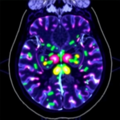
Technological advancements in imaging and molecular profiling were instrumental in delineating the complex EV content and their cellular impacts. Fluorescence microscopy, flow cytometry, and proteomic analyses collectively validated the presence of functional cytokines within vesicles and mapped their intracellular trafficking routes in melanoma cells. This comprehensive approach underscores the power of integrative methodologies in cancer research.
The inflammatory cycle perpetuated by M1 macrophage-derived EVs creates a microenvironment conducive to tumor survival and expansion, exemplifying how immune cells may inadvertently support malignancies. This paradigm-shifting discovery enhances the understanding of tumor immunology and highlights extracellular vesicles as both biomarkers and therapeutic targets for cancer intervention.
The research was spearheaded by Doctoral Researcher Kaisa Mäki-Mantila and led by Associate Professor Sanna Pasonen-Seppänen at the University of Eastern Finland’s Institute of Biomedicine. The multi-institutional support from prominent cancer research foundations reflects the study’s significance and potential translational impact in oncology.
In summary, this groundbreaking investigation reveals a sophisticated mechanism by which pro-inflammatory M1 macrophages promote melanoma progression via the secretion of extracellular vesicles laden with cytokines that activate NF-κB signaling in tumor cells. By perpetuating a pro-inflammatory and invasive tumor microenvironment, these vesicles enable cancer cells to thrive and spread. Unraveling this interplay paves the way for innovative therapeutic strategies aimed at disrupting this deleterious cellular communication network to improve melanoma patient prognosis.
Subject of Research: Interaction between pro-inflammatory M1 macrophage-derived extracellular vesicles and melanoma cell behavior.
Article Title: Extracellular vesicles derived from pro-inflammatory M1 macrophages induce an inflammatory and invasive phenotype in melanoma cells.
News Publication Date: 3 December 2025
Web References:
10.1186/s12964-025-02571-8
Image Credits: Kaisa Mäki-Mantila
Keywords:
Melanoma, extracellular vesicles, M1 macrophages, inflammation, NF-κB signaling, cytokines, tumor microenvironment, TNFα, IL-1β, cancer invasion, immune cell communication, tumor progression