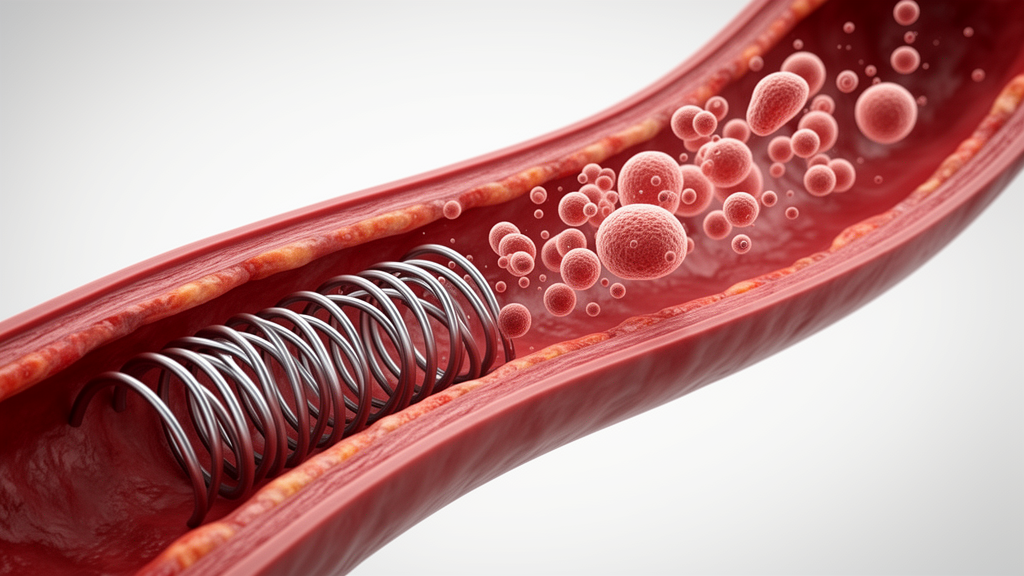
Higher preprocedural monocyte distribution width is significantly associated with the development of in-stent restenosis among patients undergoing drug-eluting stent implantation for coronary artery disease, according to a retrospective cohort study conducted by researchers at Nanjing Medical University in Nanjing, China. The investigation, led by Qian Ma, Ziwen Song, Tingting Huang, and corresponding author Runfeng Sun, evaluated whether circulating monocyte cell size variability measured before intervention could serve as an independent marker for post-procedural vascular narrowing. While drug-eluting stents have notably enhanced therapeutic outcomes for individuals with coronary artery disease, in-stent restenosis persists as an important clinical complication following percutaneous coronary intervention, highlighting an ongoing need to clarify preprocedural risk factors.
To explore this clinical question, the investigative team designed a single-center retrospective cohort study involving consecutive coronary artery disease patients who underwent successful drug-eluting stent placement between April 2022 and March 2025. Across the entire study period, 1,035 individuals met the baseline eligibility criteria for inclusion. Among this initial cohort, 982 patients underwent definitive post-procedural vascular imaging assessment to verify the presence or absence of in-stent restenosis between 6 and 14 months after their percutaneous coronary intervention. These 982 fully assessed individuals constituted the primary analytical cohort analyzed by the researchers.
Follow-up vascular evaluations were performed using either invasive coronary angiography or noninvasive coronary computed tomography angiography to objectively confirm lumen status. In the primary analysis of 982 patients, the clinicians identified 89 confirmed in-stent restenosis events, reflecting an overall incidence rate of 9.1 percent during the follow-up window. When baseline hematologic parameters were examined, patients who subsequently developed in-stent restenosis exhibited significantly higher preprocedural monocyte distribution width values than those who remained free of the complication. Specifically, the restenosis group had a median preprocedural width of 23.1 femtoliters with an interquartile range of 20.2 to 26.5 femtoliters, compared to a median of 20.9 femtoliters with an interquartile range of 18.7 to 23.6 femtoliters among patients without restenosis, representing a statistically significant difference with a P value of less than 0.001.
The researchers applied restricted cubic spline analysis to characterize the underlying shape of the relationship between preprocedural monocyte distribution width and in-stent restenosis risk across the patient population. This modeling demonstrated a statistically significant overall association between the biomarker and restenosis events, with a P value of less than 0.001. Concurrently, the spline analysis yielded a P value for nonlinearity of 0.417, demonstrating that the observed relationship did not deviate significantly from linearity across the spectrum of measured values.
To determine whether this observed relationship was independent of established clinical and procedural influences, the investigators constructed multivariable logistic regression models. These models adjusted for a comprehensive set of prespecified clinical and anatomical covariates, specifically patient age, the presence of diabetes mellitus, coronary lesion complexity, total implanted stent length, average stent diameter, and low-density lipoprotein cholesterol concentrations. Following multivariable adjustment, preprocedural monocyte distribution width remained an independent factor associated with higher odds of restenosis, with each 1-femtoliter elevation corresponding to a 17 percent increase in the odds of in-stent restenosis, supported by an odds ratio of 1.17, a 95 percent confidence interval spanning 1.07 to 1.28, and a P value of less than 0.001.
The consistency of this statistical association was further evaluated in designated subgroup and sensitivity assessments. Among a subset of 742 patients who underwent follow-up assessment using invasive coronary angiography as their primary imaging modality, multivariable regression yielded comparable results, showing an odds ratio of 1.15 with a 95 percent confidence interval of 1.05 to 1.27 and a P value of 0.003 for each 1-femtoliter increase. Furthermore, the statistical association between baseline monocyte distribution width and in-stent restenosis persisted when models were subjected to additional sensitivity analyses that adjusted for other inflammatory parameters, including absolute circulating monocyte counts and high-sensitivity C-reactive protein levels.
In interpreting their findings, the study authors suggested that elevated preprocedural monocyte distribution width may reflect a monocyte-related inflammatory phenotype that correlates with increased biological susceptibility to in-stent restenosis following drug-eluting stent implantation. However, the authors explicitly emphasized several critical limitations and uncertainties regarding practical implementation. They noted that the biomarker’s incremental predictive value over conventional risk assessment, its optimal diagnostic and clinical threshold, and its actual clinical utility remain unestablished. Consequently, prospective multicenter studies conducted in independent patient populations are required before monocyte distribution width can be considered for routine clinical evaluation.
Subject of Research: Association between preprocedural monocyte distribution width and in-stent restenosis after drug-eluting stent implantation
Article Title: Association of preprocedural monocyte distribution width with in-stent restenosis after drug-eluting stent implantation: a retrospective cohort study
Article References: Ma, Q., Song, Z., Huang, T., & Sun, R. (2026). Association of preprocedural monocyte distribution width with in-stent restenosis after drug-eluting stent implantation: a retrospective cohort study. Frontiers in Cardiovascular Medicine, 13, Article 2014164. https://doi.org/10.3389/fcvm.2026.2014164
Image Credits: AI Generated
DOI: 10.3389/fcvm.2026.2014164
Keywords: monocyte distribution width, in-stent restenosis, drug-eluting stents, percutaneous coronary intervention, coronary artery disease, inflammatory biomarkers, cardiovascular medicine, coronary angiography, coronary computed tomography angiography, retrospective cohort study, multivariable logistic regression, restricted cubic spline, high-sensitivity C-reactive protein, absolute monocyte count, low-density lipoprotein cholesterol, stent length, stent diameter, Nanjing Medical University
News Source: Ophelia Keating. (November 18, 2026). Preprocedural Monocyte Distribution Width Linked to Stent Restenosis in Retrospective Study. Scienmag.