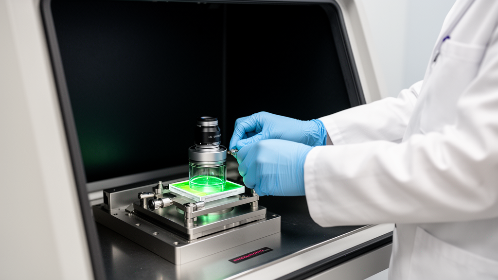
Preclinical optical imaging techniques, notably bioluminescence and fluorescence imaging, provide indispensable non-invasive windows into cellular and molecular dynamics within living animal models, yet their quantitative reliability remains heavily constrained by biochemical, host, and physical artifacts. While modalities such as magnetic resonance imaging and computed tomography offer high anatomical resolution at the cost of molecular sensitivity or scan duration, optical systems capture non-ionizing light spanning ultraviolet to near-infrared wavelengths with nanometer-scale spatial resolution and nanomolar detection limits. However, the apparent operational simplicity of optical image acquisition frequently obscures complex physical and physiological interferences that compromise experimental reproducibility if researchers fail to account for tissue optics, probe kinetics, and microenvironmental factors.
Bioluminescence imaging relies fundamentally on the enzymatic conversion of a substrate by luciferase enzymes engineered into target cells or pathogens to generate light in the presence of oxygen, adenosine triphosphate, and magnesium ions. In the canonical North American firefly system, luciferase catalyzes the oxidation of D-luciferin to produce oxyluciferin, carbon dioxide, adenosine monophosphate, pyrophosphate, and light peaking at approximately 560 nanometers. Because the initial adenylation step requires adenosine triphosphate to form a luciferyl-adenylate intermediate that subsequently reacts with molecular oxygen to generate a transient dioxetanone structure, bioluminescence serves as an intrinsic reporter of metabolic activity and cell viability. Nevertheless, this direct dependence on physiological cofactors introduces critical vulnerabilities, as tissue hypoxia or metabolic exhaustion within desmoplastic tumor cores can dramatically suppress photon emission independently of actual viable cell numbers, creating misleading readouts of treatment response.
A critical determinant of quantitative accuracy in bioluminescence imaging is maintaining absolute substrate saturation across all target populations. In animal models, D-luciferin is conventionally administered at a standardized dosage of 150 milligrams per kilogram of body weight to saturate the luciferase enzyme fully. If substrate delivery drops below saturating thresholds, photon emission ceases to correlate linearly with enzyme concentration or cell number, conflating reduced substrate availability with biological decline. Biodistribution disparities across divergent tissue beds further compound this challenge; for example, D-luciferin exhibits poor permeability across the blood-brain barrier, making the quantitative monitoring of intracranial glioblastoma highly dependent on barrier breakdown rather than purely reflecting tumor proliferation. Under such restrictive delivery conditions, extraneous pharmacological interactions can also induce severe artifacts, as demonstrated when cyclodextrin-based formulations encapsulate D-luciferin and suppress bioluminescent emission without altering true tumor growth.
These substrate delivery barriers become acutely evident when applying bioluminescence imaging to fungal pathogen models, where complex cell walls and protective extracellular biofilms severely impede substrate uptake. In fungal biofilms, the dense matrix not only restricts the diffusion of D-luciferin but also absorbs and scatters emitted photons. Experimental attempts to overcome this limitation by escalating substrate concentrations up to 4000 micrograms per gram of body weight in Galleria mellonella larvae—more than 25 times the recommended dosage—triggered overt host toxicity and disrupted pharmacokinetics without achieving uniform tissue saturation. To address these limitations, synthetic and engineered bioluminescent systems have been developed, including NanoLuc from Oplophorus gracilirostris, which operates with furimazine to generate light over 150-fold brighter than firefly luciferase, and the red-shifted Akaluc/AkaLumine pairing, which emits near-infrared light capable of penetrating deep mammalian tissues down to single-cell detection limits in pulmonary metastasis models.
Host physiology and physical tissue architecture impose additional physical boundaries on bioluminescent signal fidelity across living organisms. Photons traversing biological tissue experience extensive wavelength-dependent absorption by endogenous chromophores, primarily hemoglobin, water, and lipids, alongside extensive scattering by dense cellular interfaces and extracellular matrix networks. Hypoperfusion reduces luciferin delivery to ischemic lesions, while dense fibrotic matrices impair passive diffusion. Ambient and core body temperature fluctuations also introduce signal instability, given that luciferase enzymatic reaction rates peak at specific thermal optima before suffering thermal denaturation. Furthermore, systemic administration of substrates such as AkaLumine can induce substantial liver-specific background luminescence due to non-target metabolism by hepatic enzymes, obscuring target signals in abdominal regions unless researchers employ rigorous controls and signal deconvolution algorithms.
The mathematical processing of background noise represents another pivotal factor in preventing false findings during bioluminescence analysis, particularly when recording faint signals. Bioluminescent signals frequently span multiple orders of magnitude, requiring representation on logarithmic scales; however, standard background subtraction can yield zero or negative net values that are mathematically undefined under logarithmic transformation. When true biological emission approaches ambient background thresholds, subtracting background values amplifies experimental variability and generates artificial fold differences. Normalizing data by dividing signal intensity by background levels preserves mathematical proportionality, ensures that normalized values remain positive, directly reflects true signal-to-noise ratios, and significantly enhances the statistical reproducibility of comparative longitudinal measurements across cohorts.
Preclinical whole-body fluorescence imaging operates via distinct physical mechanisms, relying on external light excitation of exogenous fluorophores, fluorescent proteins, or nanoprobes rather than an internal enzymatic reaction. Modern fluorescence detection systems achieve detection sensitivities spanning picomolar to nanomolar concentrations, offering versatile multiplexing capabilities where distinct fluorophores allow simultaneous tracking of multiple cellular targets or functional processes such as caspase activation and immune cell recruitment. However, planar two-dimensional fluorescence imaging provides a spatial resolution limited to roughly 1 to 2 millimeters in small animals, and photon scattering across tissue layers causes rapid loss of spatial detail as depth increases. Optical absorption by blood hemoglobin and scattering by animal hair or skin pigmentation further distort signal intensity, necessitating reproducible shaving protocols with depilation creams and precise spectral filtering.
Autofluorescence from endogenous biological components represents the primary confounding factor in preclinical fluorescence imaging, particularly within the visible light spectrum. Structural proteins such as collagen and elastin, alongside metabolic cofactors like flavins and porphyrins, emit broad fluorescent backgrounds upon excitation with ultraviolet or blue light. Diet-derived fluorescent compounds, including chlorophyll present in standard animal feed, accumulate within the gastrointestinal tract and generate intense abdominal background signals that necessitate dietary fasting prior to imaging. Moving into the near-infrared II window spanning 1000 to 1700 nanometers markedly reduces tissue autofluorescence and photon scattering, enabling imaging penetration depths of up to several centimeters with enhanced signal-to-noise ratios, reduced phototoxicity, and minimal photobleaching during prolonged dynamic vascular examinations.
Advanced hybrid instrumentation platforms have progressively emerged to resolve the depth and quantification deficiencies inherent to planar optical imaging modalities. Systems combining fluorescence emission computed tomography with micro-computed tomography utilize 48-detector rings and anatomical cross-sectional reconstructions to account for tissue-specific absorption coefficients and organ boundaries, translating detected surface photons into accurate three-dimensional source concentrations. Dual-modality approaches merging bioluminescence and fluorescence capitalize on the high signal-to-noise ratio and low background of luciferase reporters for early lesion detection while deploying targeted fluorophores for high-resolution anatomical localization. To avoid cross-talk, dual-imaging protocols require performing fluorescence scans prior to substrate injection or selecting spectrally separated reporters like near-infrared fluorescent proteins to prevent uncontrolled bioluminescence resonance energy transfer.
Beyond physical and optical parameters, the biological consequences of introducing foreign optical reporters into host organisms require vigilant experimental controls. High-level expression of luciferase under strong promoters can trigger unfolded protein responses and metabolic stress, while continuous ATP consumption during frequent imaging sessions risks depleting cellular energy reserves and elevating reactive oxygen species. Because luciferases originate from non-mammalian organisms such as insects and marine coelenterates, they frequently evoke neutralizing antibody responses and immune inflammation in immunocompetent animals, progressively attenuating longitudinal signals. By establishing standardized substrate delivery protocols, applying background division, accounting for tissue attenuation, and incorporating anatomical priors, researchers can safeguard optical imaging as a robust, non-invasive standard for monitoring disease progression and therapeutic efficacy.
Subject of Research: Optimization guidelines and physical limitations of preclinical optical imaging techniques
Article Title: Optical imaging in biomedical research: guidelines and practical insights
Article References: Chu, T., Zheng, X., Manshian, B. B., & Soenen, S. J. (2025). Optical imaging in biomedical research: guidelines and practical insights. Health Nanotechnology, 1(1), Article 4. https://doi.org/10.1186/s44301-025-00004-6
Image Credits: AI Generated
DOI: 10.1186/s44301-025-00004-6
Keywords: bioluminescence imaging, fluorescence imaging, preclinical imaging, firefly luciferase, D-luciferin, NanoLuc, AkaLumine, near-infrared fluorescence, tissue autofluorescence, signal-to-noise ratio, spectral unmixing, FLECT-CT, molecular imaging, subcellular imaging, preclinical oncology, optical tomography
News Source: Ophelia Keating. (November 19, 2026). Preclinical Optical Imaging Demands Rigorous Standardization to Overcome Severe Quantification Artifacts. Scienmag.