When a severe stroke strikes, the ambulance crew faces a decision that can shape the rest of a patient’s life: drive to the nearest hospital, or bypass it for a distant comprehensive stroke center equipped to pull a clot out of the brain. A new analysis published in Frontiers in Neurology suggests that a portable, non-imaging ultrasound device that measures brain perfusion could help make that call more reliable. In a secondary analysis of the SONAS trial, researchers from the University of Regensburg and the University of California, Los Angeles, found that pairing the device, known as SONAS brain perfusion ultrasound, with established prehospital stroke scales allowed nearly every patient in their small cohort to be sorted confidently into one of three categories: highly probable large vessel occlusion, highly probable non-occlusion, or unclear.
The stakes of this decision are considerable. Stroke is the second leading cause of death worldwide, and while intravenous thrombolysis and mechanical thrombectomy have transformed outcomes, both treatments are fiercely time-dependent. Mechanical thrombectomy can restore blood flow in patients whose large brain arteries are blocked, but comprehensive stroke centers capable of performing the procedure are unevenly distributed, particularly in rural regions. When a patient with a large vessel occlusion is first taken to a smaller primary stroke center and then transferred for thrombectomy, the so-called drip-and-ship pathway, the secondary transport consumes precious time and can worsen prognosis. Conversely, sending every suspected stroke patient directly to a comprehensive center risks overloading those centers and delaying thrombolysis for patients who do not need it.
To guide this triage, clinicians have developed a host of prehospital stroke scales, bedside checklists that score facial weakness, arm drift, gaze palsy, language disturbance, and other deficits to estimate the likelihood of a large vessel occlusion. The problem, as the Regensburg team notes, is that no scale achieves both high sensitivity and high specificity. Even after a negative test, a large vessel occlusion may still be present with a probability of at least ten percent. When prehospital scores alone are used to decide on a target hospital, roughly half of patients are inappropriately diverted to a comprehensive stroke center, which can cause harm by delaying intravenous thrombolysis. Guidelines worldwide recommend using stroke scales in the field, but none endorse a specific scale as clearly superior.
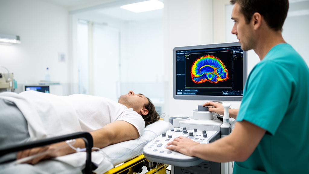
The SONAS device represents a different approach. It is a battery-powered, non-imaging ultrasound system weighing just 2.2 kilograms, built around a transducer headset that sits over the patient’s temporal skull windows like a pair of headphones. After an injection of an approved ultrasound contrast agent, the device tracks how quickly the contrast washes into each hemisphere of the brain, generating time-intensity curves that reflect perfusion. The time between injection and peak signal, the time to peak, is compared between hemispheres. A blockage of the middle cerebral artery, the vessel most commonly implicated in large vessel occlusion stroke, delays or abolishes contrast inflow on the affected side, producing a measurable asymmetry. Because the measurement is simple and simultaneous across both hemispheres, the developers envision it being applied by paramedics with telemedical support from stroke specialists.
In the original SONAS study, conducted at Regensburg and registered in the European clinical trials register, the device was tested in twenty patients with acute ischemic stroke, all of whom also underwent computed tomography perfusion imaging as the reference standard. Seventeen of the twenty patients, or eighty-five percent, had a confirmed middle cerebral artery occlusion. Fourteen patients were female, and ages ranged from forty-four to ninety-one with an average of 72.8 years. The ultrasound assessment could not be completed in two patients because of poor data quality, typically stemming from inadequate temporal bone windows or unfavorable headset placement. Among the eighteen evaluable datasets, the device correctly identified fourteen patients with occlusion and two without, misclassifying one patient in each direction, corresponding to a sensitivity of ninety-three percent and a specificity of sixty-six percent.
For the new analysis, the researchers took the detailed neurological examinations recorded in the SONAS trial and retrospectively calculated sixteen different prehospital stroke scores for each patient, using the published cutoff values that yielded the best sensitivity and specificity. The best performers were CG-FAST and G-FAST, which at a cutoff between two and three missed a large vessel occlusion in only a single patient while correctly classifying all others, yielding a sensitivity of ninety-four percent and a specificity of one hundred percent. The RACE score, at a cutoff between four and five, missed two patients, giving a sensitivity of eighty-eight percent and again perfect specificity. Notably, the full NIH Stroke Scale, a hospital-based severity measure rather than a field tool, would have classified every patient correctly at a cutoff between seven and eight, though the authors caution this reflects the small, pre-selected cohort.
The heart of the study lies in what happened when the ultrasound and the scores were combined. The researchers modeled several scenarios. If both methods agreed the patient had an occlusion, the patient was classified as positive; if both agreed there was none, the classification was negative. When the two methods disagreed, the team explored classifying the patient either as an occlusion, which favors sensitivity, or as a non-occlusion, which favors specificity. In the sensitivity-favoring scenario with CG-FAST, combined sensitivity rose to one hundred percent while specificity fell to sixty-six percent; with RACE, sensitivity likewise reached one hundred percent but specificity dropped to thirty-three percent. In the specificity-favoring scenario, results remained nearly identical to the scores alone. The combination, in other words, does not automatically improve accuracy but allows clinicians to tune the trade-off between catching every occlusion and avoiding unnecessary diversions.
The most striking result emerged from a three-way classification. Patients in whom both the ultrasound and the score agreed were labeled clear occlusion or clearly no occlusion; those with contradicting results were labeled unclear. This scheme classified every patient in the cohort correctly whenever a confident call was made: thirteen patients were jointly identified as having an occlusion and one or two as not, all correctly. Crucially, the unclear group captured every single patient who had been misclassified by either method alone, four patients using RACE and three using CG-FAST, of whom two in each group truly had an occlusion. The authors argue this makes the combined approach highly effective at flagging uncertain cases, which could then be routed based on other clinical factors and local circumstances rather than a potentially wrong confident answer.
The analysis also probed the other great diagnostic fork in prehospital stroke care: distinguishing ischemic stroke from intracerebral hemorrhage. The SONAS trial excluded hemorrhage, but the team applied a validated prehospital intracerebral hemorrhage score, developed in the Berlin mobile stroke unit project, to the twenty ischemic patients. The score incorrectly flagged three patients, fifteen percent, as likely hemorrhage. In all three, both the SONAS ultrasound and perfusion CT detected significant perfusion deficits consistent with a large vessel occlusion. The authors note this misclassification rate mirrors the RACECAT trial, in which twenty percent of patients redirected to comprehensive centers for suspected occlusion actually had hemorrhage, a finding with real consequences since the INTERACT-4 trial suggests urgent blood-pressure lowering may help hemorrhage patients but harm those with ischemic stroke.
The authors are careful about the limits of their work. The cohort comprised just twenty highly selected patients, examined by trained stroke neurologists rather than paramedics, without randomization or blinding, and excluding stroke mimics, hemorrhage, and subarachnoid hemorrhage. Validation in unselected stroke-call patients in the field, applied by paramedics with or without telemedical support, and comparison with other point-of-care diagnostics on sensitivity, speed, and cost remain essential. If those hurdles are cleared, the implications are tantalizing: high-confidence prehospital identification of ischemic stroke could enable prehospital thrombolysis with tenecteplase, serum biomarkers such as GFAP could exclude hemorrhage, confirmed occlusion patients could go directly to the angiography suite, and blood pressure and anticoagulation could be managed en route. Technical refinements to detect posterior circulation strokes, absent from this study, are already underway. For now, the message is measured: a two-kilogram ultrasound headset, paired with a bedside checklist, may one day give ambulance crews the diagnostic confidence they currently lack.
Subject of Research: Combining portable brain perfusion ultrasound with prehospital stroke scales to detect large vessel occlusion in acute stroke
Article Title: Added value of brain perfusion ultrasound to prehospital stroke scales-a secondary analysis of the SONAS trial
Article References: Birner, N., Wilfling, S., Kilic, M., Wendl, C., Molaie, A. M., Linker, R. A., Liebeskind, D. S., & Schlachetzki, F. (2026). Added value of brain perfusion ultrasound to prehospital stroke scales-a secondary analysis of the SONAS trial. Frontiers in Neurology, 17, Article 1924664. https://doi.org/10.3389/fneur.2026.1924664
Image Credits: AI Generated
DOI: 10.3389/fneur.2026.1924664
Keywords: stroke, large vessel occlusion, brain perfusion ultrasound, SONAS, prehospital triage, mechanical thrombectomy, CG-FAST, RACE score, intracerebral hemorrhage, telemedicine, emergency medicine, diagnostics
News Source: Cassandra Pierce. (October 24, 2026). Portable Brain Perfusion Ultrasound Could Sharpen Prehospital Stroke Triage, Small Trial Analysis Suggests. Scienmag.