• HOME
  • NEWS
  • EXPLORE
    • CAREER
      • Companies
      • Jobs
    • EVENTS
    • iGEM
      • News
      • Team
    • PHOTOS
    • VIDEO
    • WIKI
  • BLOG
  • COMMUNITY
    • FACEBOOK
    • INSTAGRAM
    • TWITTER
Tuesday, October 6, 2026
BIOENGINEER.ORG
No Result
View All Result
  • Login
  • HOME
  • NEWS
  • EXPLORE
    • CAREER
      • Companies
      • Jobs
        • Lecturer
        • PhD Studentship
        • Postdoc
        • Research Assistant
    • EVENTS
    • iGEM
      • News
      • Team
    • PHOTOS
    • VIDEO
    • WIKI
  • BLOG
  • COMMUNITY
    • FACEBOOK
    • INSTAGRAM
    • TWITTER
  • HOME
  • NEWS
  • EXPLORE
    • CAREER
      • Companies
      • Jobs
        • Lecturer
        • PhD Studentship
        • Postdoc
        • Research Assistant
    • EVENTS
    • iGEM
      • News
      • Team
    • PHOTOS
    • VIDEO
    • WIKI
  • BLOG
  • COMMUNITY
    • FACEBOOK
    • INSTAGRAM
    • TWITTER
No Result
View All Result
Bioengineer.org
No Result
View All Result
Home NEWS Science News Chemistry

PLUS takes 3D ultrasound images of solids

Bioengineer by Bioengineer
September 25, 2020
in Chemistry
Reading Time: 3 mins read
0
IMAGE
Share on FacebookShare on TwitterShare on LinkedinShare on RedditShare on Telegram

IMAGE

Credit: Yoshikazu Ohara, Tohoku University

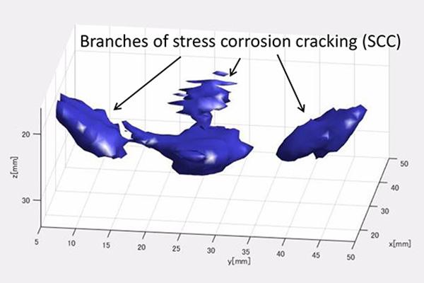
A new system, developed by Tohoku University researchers in Japan in collaboration with Los Alamos National Laboratory in the US, takes 3D images that can detect defects in metallic structures. The approach was published in the journal Applied Physics Letters and could enhance safety in power plants and airplanes.

Yoshikazu Ohara and colleagues at Tohoku University use non-destructive techniques to study structures, and wanted to find a way to produce 3D images of structural defects. They developed a new technology, called the piezoelectric and laser ultrasonic system (PLUS), that combines the strengths of two different devices to produce high-resolution 3D images of defects in metallic structures.

“We believe that PLUS will pave the way for accurate evaluation of material strength, the identification of defects, and finding out how defects initially started to form,” says Ohara.

Currently available ‘ultrasonic phased arrays’ are a powerful tool for imaging internal defects in solids, but only in two dimensions. These devices are made of a piezoelectric one-dimensional array transducer with a limited number of individual elements–up to 128. Electrical pulses in the piezoelectric elements are converted to a mechanical vibration that emits ultrasonic waves into the material under investigation. Ultrasonic waves are reflected back from internal defects and converted into electric signals that can be translated into a 2D image.

In PLUS, the waves generated in a material from a piezoelectric transducer with a single element are received by a laser Doppler vibrometer, which moves around the material’s surface to get a good 2D scan of the area. As a result of this process, it receives the scattered and reflected waves at a much larger number of ‘points’ than those that can be received by a piezoelectric array transducer. The information received by the laser Doppler vibrometer is transmitted by an oscilloscope to a computer, where it is processed by an imaging algorithm and converted into a 3D image.

“Ultrasonic phased arrays, which are on the cutting-edge of ultrasonic inspection, can only provide 2D images because of their limited number of elements,” says Ohara. “PLUS makes it possible to have thousands of elements as a result of incorporating the 2D scan of a laser Doppler vibrometer in place of a piezoelectric array transducer.”

Although tested only on defects in metallic materials, Ohara says their technology can be applied to other materials, including concrete and rock, simply by changing the phased array transmitter to one that emits a different range of ultrasound frequencies.

One drawback is the long data acquisition and processing time, which takes several hours. However, this can be shortened by adopting a high-speed analog-to-digital converter in place of the oscilloscope, using a more sensitive laser Doppler vibrometer, utilizing different imaging algorithms, and employing a graphical processing unit.

###

Media Contact
Yoshikazu Ohara
[email protected]

Original Source

https://www.tohoku.ac.jp/en/press/plus_3d_images_2020.html

Related Journal Article

http://dx.doi.org/10.1063/5.0021282

Tags: Chemistry/Physics/Materials SciencesCivil EngineeringMaterials
Share12Tweet8Share2ShareShareShare2

Related Posts

Water-Repelling MOF Turns Humid Flue Gas Into an Advantage for Carbon Capture

Water-Repelling MOF Turns Humid Flue Gas Into an Advantage for Carbon Capture

October 6, 2026
Handheld NIR Scanner and Bilinear Surface Model Crack Rapid Gluten Quality Testing in Whole Wheat Flour

Handheld NIR Scanner and Bilinear Surface Model Crack Rapid Gluten Quality Testing in Whole Wheat Flour

October 6, 2026

Dunes Follow a Hidden Physics: Young Coastal Dune Landscapes Self-Organize Like Phase-Separating Fluids

October 6, 2026

Fungus Hidden Inside Ginger Yields Fatty Acid That Kills Malaria Mosquito Larvae

October 6, 2026
Please login to join discussion

POPULAR NEWS

  • Alloys That Shrink Their Own Grains: New PIX Mechanism Refines Metals With Heat Alone

    Alloys That Shrink Their Own Grains: New PIX Mechanism Refines Metals With Heat Alone

    29 shares
    Share 12 Tweet 7
  • Endurance Exercise Reshapes the Liver in Males and Females Through Distinct Molecular Routes

    29 shares
    Share 12 Tweet 7
  • Single Transcription Factor PU.1 Rapidly Converts Fibroblasts into Macrophage-Lineage Cells

    29 shares
    Share 12 Tweet 7
  • New Scale Measures How Ready Nurse Educators Really Are for the AI Era

    29 shares
    Share 12 Tweet 7

About

We bring you the latest biotechnology news from best research centers and universities around the world. Check our website.

Follow us

Recent News

Alloys That Shrink Their Own Grains: New PIX Mechanism Refines Metals With Heat Alone

Endurance Exercise Reshapes the Liver in Males and Females Through Distinct Molecular Routes

Single Transcription Factor PU.1 Rapidly Converts Fibroblasts into Macrophage-Lineage Cells

Subscribe to Blog via Email

Success! An email was just sent to confirm your subscription. Please find the email now and click 'Confirm' to start subscribing.

Join 85 other subscribers
  • Contact Us

Bioengineer.org © Copyright 2023 All Rights Reserved.

Welcome Back!

Login to your account below

Forgotten Password?

Retrieve your password

Please enter your username or email address to reset your password.

Log In
No Result
View All Result
  • Homepages
    • Home Page 1
    • Home Page 2
  • News
  • National
  • Business
  • Health
  • Lifestyle
  • Science

Bioengineer.org © Copyright 2023 All Rights Reserved.